VitroVivo Special Staining Services – Expert Tissue Staining for Research & Diagnostics
VitroVivo Biotech provides special staining services designed to enhance tissue visualization beyond standard Hematoxylin and Eosin (H&E) staining. Special stains help identify specific tissue structures, cellular components, microorganisms, metals, salts, and more that are not easily detected by routine staining.
What Are Special Stains?
🔬 Beyond H&E Staining – Special stains use advanced techniques to differentiate specific tissue components.
🧪 Precision & Customization – Essential for research, pathology, and clinical diagnostics.
🧬 Diverse Applications – Supports studies in histology, microbiology, and molecular biology.
Types of Special Stains
Special stains can be categorized into different groups based on their application:
✔ Histological Stains – Identify structural components within tissue.
✔ Microbial Stains – Detect bacteria, fungi, and other microorganisms.
✔ Histochemical Stains – Highlight specific biomolecules, metals, and deposits.
✔ Advanced Stains – Includes immunohistochemistry (IHC) and in situ hybridization (ISH) for protein and DNA/RNA analysis.
VitroVivo Special Staining Services
We offer a range of high-quality special stains using standardized protocols:
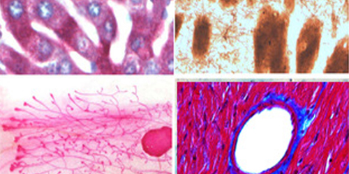
✅ Mammary Gland Whole Mount Staining
✅ Alcian Blue Hematoxylin/Orange G Staining
✅ Oil Red O Staining (for lipid detection)
✅ Modified Gomori’s Trichrome Staining (for muscle and connective tissues)
✅ Bielschowsky’s Silver Staining (for neural structures)
✅ Masson’s Trichrome Staining (for collagen fibers)
✅ Picro-Sirius Red Staining (for collagen analysis)
✅ Reticulum Staining (for reticular fibers)
✅ Custom Special Stains – Available upon request.
DIY Special Staining Kits
Prefer to perform staining in your own lab? VitroVivo offers special stain kits for self-use, ensuring high-quality and reproducible results.
📩 Contact us at service@vitrovivo.com for inquiries, pricing, or custom staining solutions tailored to your research needs!
Frequently Asked Questions (FAQ)
FAQ 1. What is a special stain?
Any stain, other than an H&E stain, is classified as a special stain. The common special stains include mammary gland whole mount stain, alcian blue hematoxylin-orange G stain, alcian blue stain, alcian blue – PAS stain, oil red O stain, Nissl stain, Bielschowsky’s silver stain and Masson’s trichrome stain, etc. For more information, please visit our website page of Histochemical Stain Kits and Image Gallery.
FAQ 2. How do I choose VitroVivo special staining services?
This table gives you a guidance of the choice of special staining methods or kits (if you want to perform staining by your self). If you can not find the staining methods from this table, please send your email to service@vitrovivo.com for inquiry, our service team will get back to you as soon as possible.
| Product Name | SKU# | Visualization for | Typical Results |
| Hematoxylin and Eosin Kit | VB-3000 | General morphology of tissue and cell |
|
| Mammary Gland Whole Mount Stain Kit | VB-3001 | Wholemount staining of mouse mammary glands |
|
| Alcian Blue Hematoxylin-Orange G Stain Kit | VB-3002 | Differentiate cartilage, mature bone, and immature bone found in various stages of endochondral ossification and fracture callus |
|
| Alcian Blue Stain Kit | VB-3003 | Tisssue mucosubstances |
|
| PAS Stain Kit | VB-3004 | Glycogen, mucin, and fungi |
|
| Alcian Blue – PAS Stain Kit | VB-3005 | Acidic and neutral mucins as well as mixtures of acidic and neutral mucins |
|
| Luxol Fast Blue Stain Kit | VB-3006 | Myelin including phospholipids and neurons |
|
| Oil Red O Stain Kit | VB-3007 | lipid and fat staining on formalin fixed frozen sections |
|
| Alizarin Red Stain Kit | VB-3008 | Calcium on tissue sections |
|
| Prussian Blue Stain Kit | VB-3009 | Ferric iron on tissue sections |
|
| Nissl Stain Kit | VB-3010 | Neuron Nissl body |
|
| Congo Red Amyloid Stain Kit | VB-3011 | Amyloid deposits |
|
| Sudan Black B Lipid Stain Kit | VB-3012 | Lipid and fat |
|
| Toluidine Blue Stain Kit | VB-3013 | Mast cells |
|
| Modified Gomori’s Trichrome Stain Kit | VB-3014 | Connective fiber |
|
| Bielschowsky’s Silver Stain Kit | VB-3015 | Axons, neurofibrillary tangles and senile plaques |
|
| Masson’s Trichrome Stain Kit | VB-3016 | Collagen and mucus |
|
| Picro-Sirius Red Stain Kit | VB-3017 | Collagen fibers |
|
| Reticulum Stain Kit | VB-3018 | Reticular fibers |
|
| Verhoeff Van Gieson Elastin Stain Kit | VB-3019 | Elastic fibers |
|
| Fontana-Masson Stain Kit | VB-3020 | Melanin pigment and argentaffin granules |
|