Description
Alizarin Red Stain kit is used for identifying calcium on tissue sections. Calcium forms an alizarin red S-calcium complex in a chelation process. The reaction is not strictly specific for calcium, since magnesium, manganese, barium, strontium, and iron may interfere, but these elements usually do not occur in sufficient concentration to interfere with the staining.
Kit Contents
- Alizarin Red Solution – 250 ml
Storage Condition:
Room temperature.
The pH for kit is critical, so check pH if the solution has been stored for more than one month. Adjust the pH to 4.1~4.3 with 10% ammonium hydroxide if necessary.
Protocol
- Deparaffinize and hydrate to 70% alcohol.
- Rinse rapidly in distilled water.
- Alizarin red S solution, 30 seconds to 5 minutes, checking microscopically for an orange-red color.
- Shake off excess dye, blot sections.
- Acetone, 20 dips.
- Acetone-xylene (1:1), 20 dips.
- Clear in xylene (5min x2).
- Mount in Mount coverslip onto glass slide with VitroView™ Permanent Mounting Medium (VB-8003) or some other suitable organic mounting medium.
Results
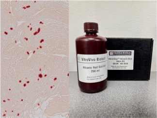
Calcium deposits: orange-red
Note: This product is intended for research purposes only. This product is not intended to be used for therapeutic or diagnostic purposes in humans or animals.
Precautions: Handle with care. Avoid contact with eyes, skin and clothing. Do not ingest. Wear gloves.
Images

Mouse aorta artery, Alizarin Red Stain |

Mouse aorta artery, Alizarin Red Stain |
User Manual and Material Safety Data Sheet (MSDS) (PDF)
Frequently Asked Questions
What does this kit detect?
Calcium deposits on tissue sections. Calcium forms an alizarin red S–calcium complex in a chelation process, and calcium deposits stain orange-red. The reaction is not strictly specific for calcium, since magnesium, manganese, barium, strontium, and iron may interfere, but these elements usually do not occur in sufficient concentration to interfere with the staining.
What sample types can I use it on?
Tissue sections. The protocol begins by deparaffinizing the sections and hydrating them to 70% alcohol.
What is in the kit?
Alizarin Red Solution, 250 ml.
How should I store it?
At room temperature. The pH of the kit is critical, so check the pH if the solution has been stored for more than one month, and adjust it to 4.1–4.3 with 10% ammonium hydroxide if necessary.
How long does the staining step take?
Stain in the Alizarin red S solution for 30 seconds to 5 minutes, checking microscopically for an orange-red color. Afterwards, shake off the excess dye, blot the sections, dip in acetone 20 times, then in acetone–xylene (1:1) 20 times, clear in xylene, and mount with VitroView™ Permanent Mounting Medium (VB-8003) or another suitable organic mounting medium.
Is this kit for diagnostic use?
No. It is intended for research purposes only, and is not intended for therapeutic or diagnostic use in humans or animals.