Description
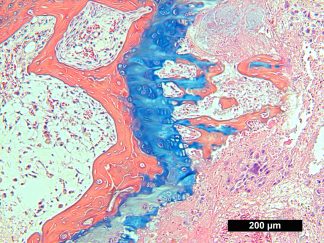
VitroView™ Alcian Blue Hematoxylin-Orange G Stain Kit is designed to stain formalin fixed, paraffin embedded tissue sections of bone and cartilage. This method can differentiate cartilage, mature bone, and immature bone found in various stages of endochondral ossification and fracture callus.
Kit Contents
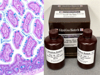
- VB-3002-1 Alcian Blue Hematoxylin—–250 ml
- VB-3002-2 Eosin/Orange G solution——250 ml
Storage Condition:
Room temperature.
Protocol
- Deparaffinize in xylene I for 6 minutes and II for 6 minutes.
- Rehydrate: ethanol 100% (2 minutes)x2; ethanol 95% (2 minutes)x2; ethanol 70% (2 minutes)x1
- Rinse in distilled water (5 minutes).
- Place in acid-alcohol (1% concentrated hydrochloric acid (HCl 37%) in 70% ethanol) for 30 seconds and drain briefly on paper towel (DO NOT RINSE)
- Place in Alcian Blue Hematoxylin for 30 minutes.
- Wash gently in distilled water until excess stain stops leaching from tissue (approximately 3 changes).
- Differentiate in acid-alcohol for 3 seconds.
- Rinse gently in distilled water with 3 changes.
- Place in 0.5% Ammonium water for 15 seconds.
- Rinse in distilled water with 2 changes.
- Place in 95% ethanol for 1 minute (DO NOT RINSE).
- Place in Eosin/Orange G for 1 min 30sec.
- Dehydrate with 3 changes of 95% Ethanol and 2 changes of 100% Ethanol (2 minute per change).
- Clear with 3 changes of xylene (5 minute per change)
- Mount coverslip onto glass slide with Permount or some other suitable organic mounting medium.
Results
- Bone: orange to red
- Activated osteocytes: bright blue pericellular ring
- Growth plate: pale blue to blue
- Cartilage: blue/purple (GAG/proteoglycan)
- Erythrocytes: bright pink
- Soft tissues (muscle, tendon, membranes): pink to red
- Bone marrow: dark blue
*Colors are more intense in Formic acid decalcified sections compared with EDTA decalcified sections.
Selected Peer-reviewed publications and preprints that have cited the use of the VitroView™ Alcian Blue Hematoxylin-Orange G Stain Kit (VB-3002)
- Isaac CV, et al.(2022). A method for the development of cranial fracture histology slides. Journal of Forensic sciences 2022, 67(5):2040-2047
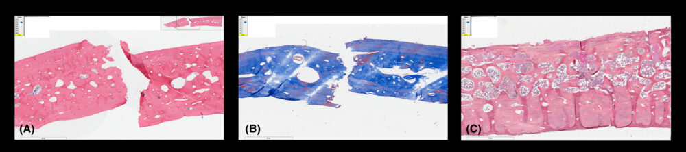
Figure 5. Common artifacts resulting from microtome sectioning. (A) H&E slide with folding along the fracture margins. (B) Trichrome slide with knife marks indicated by oblique striations. (C) AB/OG slide of a fracture in advanced healing which shows extensive wrinkling of the sample along inferior margin. 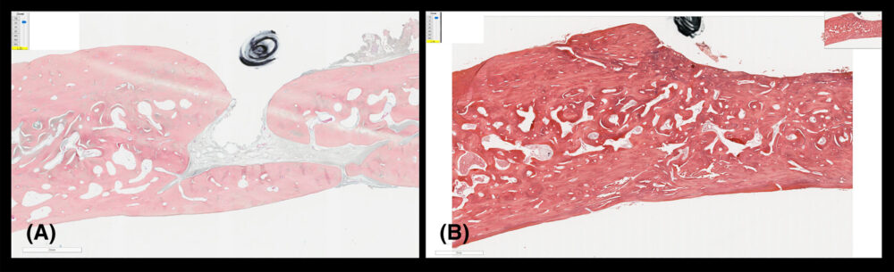
Figure 6. The variable results of AB/OG staining. (A) A washed-out AB/OG slide due to old or exhausted staining reagents. (B) A properly stained AB/OG slide with fresh reagents.
Note: This product is intended for research purposes only. This product is not intended to be used for therapeutic or diagnostic purposes in humans or animals.
Precautions: Handle with care. Avoid contact with eyes, skin and clothing. Do not ingest. Wear gloves.