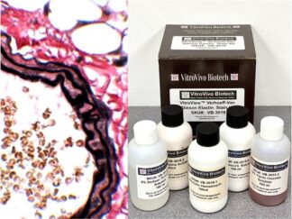
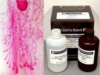
Wholemount staining of mouse mammary glands shows the structure of the whole gland and how it is altered in diseased states and in response to various hormones, or carcinogens. VitroView™ Mammary Gland Whole Mount Stain Kit is very useful for the study of mammary gland development.
Kit Contents
VB-3001-1 Carnoy’s fixative Solution—————-250ml
VB-3001-2 Carmine Alum Staining Solution——-250ml
Storage Condition
If stored at 4 oC, the kit can be used for several months. Discard when color becomes weak.
Material Needed But NOT Supplied with the Kit
Staining jar
Glass slides
Ethanol
Xylene
Mount media
Protocol
Mammary gland tissues are spread out on glass slide.
Fix in Carnoy’s fixative for 2 to 4 hours at room temperature.
Wash in 70 % Ethanol for 30 min. Change gradually to distilled water
Rinse in distilled water for 5 min.
Stain in carmine alum staining solution overnight at room temperature.
Wash in 70 % Ethanol for 15 min
Wash in 95 % Ethanol for 15 min
Wash in 100 % Ethanol for 15 min
Clear with 3 changes of xylene (10 minute per change)
Mount coverslip onto glass slide with Permount or some other suitable organic mounting medium.
Note: Carnoy’s Fixative Solution and Carmine Alum Staining Solution can be reused.
After photographic documentation the tissue can be embedded in paraffin for sectioning and conventional histological staining.
Immerse slide into xylene to remove mounting medium
Change xylene 2 times
Transfer to 1:1 xylene:paraffin (60oC)
Change xylene 2 times and embed in paraffin
Peer-Reviewed Publications Using the Mammary Gland Whole Mount Stain Kit
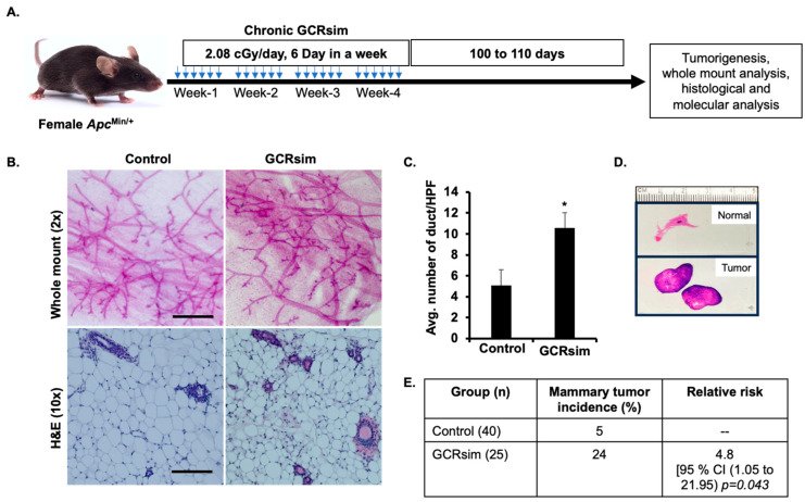
Figure 1. GCRsim exposure promotes mammary tissue overgrowth and increased duct density. (A) Schematic representation of the experimental setup depicting chronic exposure to GCRsim, using a 33-ion mixed beam to simulate the deep-space environment. Experimental animals were exposed to 2.08 cGy per day, 6 days per week, for 4 weeks, resulting in a cumulative dose of 50 cGy. (B) Representative images of mammary tissues: whole-mount micrographs (scale bar = 500 µm) show the extent of ductal branching, while H&E-stained sections (scale bar = 100 µm) highlight histological features. (C) Quantification of ductal density, expressed as the number of ducts per high-power microscopic field (HPF). (D) Representative H&E-stained normal mammary gland and tumor samples. (E) Comparison of mammary tumor incidence between control and GCRsim-exposed animals, showing increased tumor formation in the 50 cGy irradiated GCRsim group. Statistically significant difference (p < 0.05) relative to the control group is denoted by an asterisk (*).
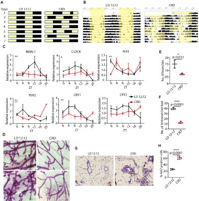
Fig. 1. CRD severely impairs rhythmicity and disrupts mammary gland morphogenesis. (A)Schematic representation of the CRD protocol. (B) FVB female mice were housed under LD 12:12 or CRD for 8 weeks. Representative actograms of LD and CRD mice. LD: 12 h light and 12 h dark; CRD: circadian rhythm disruption using jet lag protocol, represented by a shortening of the dark period by 8 h every third day. (C) Mammary glands were collected at the indicated times. Gene expression normalized to GAPDH expression was measured using quantitative real-time PCR data represented as mean ± SEM for n=3 females per time point and light condition. Rhythmicity was determined using JTK_Cycle analyses; *PJTKcycle < 0.05, **PJTKcycle <0.01, ***PJTKcycle < 0.001, and ****PJTKcycle < 0.0001. CRD disrupts branching morphogenesis in WT virgin FVB mice. (D, E, F) Representative images of branching and development of terminal ducts in LD and CRD animals are shown in LD and CRD-induced mammary glands (D, E, F), scale bar: 350 µm, (*P < 0.05, error bars indicate SEM, n = 3). (G)The effect of CRD on mammary ductal hyperplasia is shown by hematoxylin and eosin (H&E) staining of the mammary gland (G) and by using Ki67 (H) of the mammary glands. Scale bar: 100 µm.
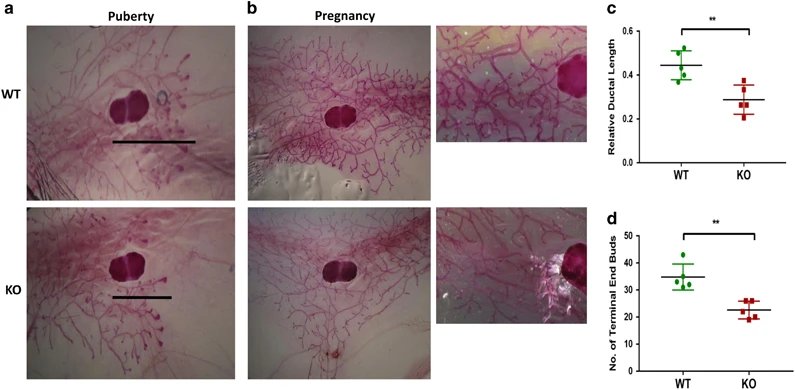
Figure 4. a2V deficiency impairs ductal elongation and bifurcation. Carmine alum staining and quantification of mammary gland whole mounts from puberty and pregnancy. (a) In pubertal mice, a2V-KO glands show delayed ductal outgrowth compared with WT. Lines represent ductal migration from the beginning of lymph node to the tip of furthest migrating duct. (b) Whole mounts from early pregnant (pregnant day 4.5) mammary gland demonstrate reduced tertiary branching and elongation in a2V-KO mice. Images were taken with x2.5 (left panels) and x20 (right panels) objective. (c) Quantification of relative ductal length was measured as a difference between whole length of the gland (from nipple end to tip of ductal tree) and ductal migration (from beginning of lymph node to the tip of ductal tree). (d) Quantification of number of TEBs (end nodes of ducts) in WT and KO mice. Data represent mean±S.E., n=6. **P≤0.01
Note
This product is intended for research purposes only. This product is not intended to be used for therapeutic or diagnostic purposes in humans or animals.
Precautions
Handle with care. Avoid contact with eyes, skin and clothing. Do not ingest. Wear gloves.
More Images From VitroVivo Biotech
FVB mouse Mammary Gland Whole Mount Staining
User Manual and Material Safety Data Sheet (MSDS) (PDF)