In Situ Detection of Cell Proliferation
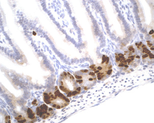
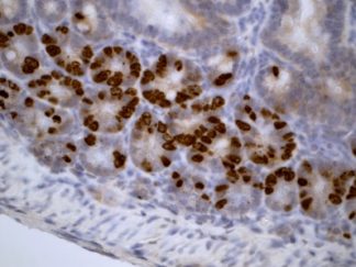
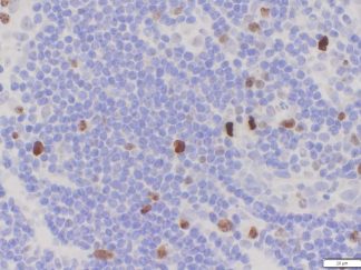
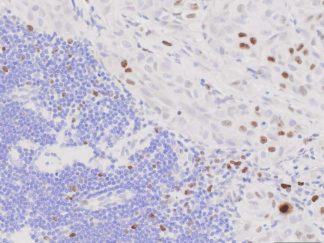
The assessment of cell proliferation in experimental animal tissues is crucial for toxicology and carcinogenesis studies, as well as for evaluating the efficacy of cytotoxic and chemopreventive drugs in cancer research. There are several methods available for detecting proliferating cells in tissue sections. The gold standard has been the in vivo labeling of DNA using a halogenated derivative of thymidine called BrdU. BrdU can be administered to laboratory animals through IP injection for pulse-label experiments or via osmotic pumps for continuous-label studies. BrdU incorporates into nuclei during the DNA synthesis phase of the cell cycle (S-phase), and its presence is detected by IHC with an anti-BrdU antibody. BrdU IHC is useful in identifying S-phase cells. Another IHC technique that has been applied in the assessment of cell proliferation is the detection of Ki-67. Ki-67 is a ubiquitous human nuclear protein expressed in G1-, S-, and G2-phases of the cell cycle, but not in the G0-phase, making it a measure of the growth fraction. Ki-67 IHC has been well-established as useful in tumor diagnostics for various malignancies.
Showing all 4 results