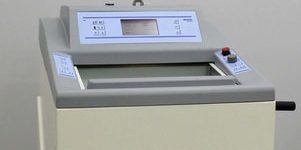
Cryotomy, frozen sectioning or cryo-sectioning is a technique that a cryotome is used to prepare thin and frozen sections for biological tissues. Frozen sections can be used for tissue analysis that allows for rapid interpretation and diagnosis of the tissue during surgery. Cryotomy can also be used in the preparation of sections containing fats and enzymes which can easily be lost in alcohol or paraffin sections.
FAQ 2. How do I prepare OCT embedding block for unfixed fresh tissue?
Flash frozen fresh tissue in OCT is a common method for frozen section preparation. It features:
| Pros | Cons |
|
|
Protocol
Place a drop of Optimal cutting temperature compound (OCT compound) in the bottom of the mold and place the tissue in the OCT. This will hold the tissue in place while you fill the mold with OCT. Just be careful to exclude large bubbles, fill the mold level full, and freeze by one of the methods below.
- Method 1:
Use dry ice in pellet form. Place a small stainless steel bowl (or Pyrex or polypropylene beaker) in the bottom of a styrofoam container and fill the space around the bowl with dry ice pellets. Place some pellets in the bowl and slowly add isopentane (2-methyl butane) or acetone. Work in a fume hood, of course, as these are flammable. When the pellets stop bubbling vigorously, the “slurry” is ready. Once you’ve filled the mold and oriented the tissue, immerse it in the liquid to freeze it.
- Method 2.
Isopentane also can be chilled in liquid nitrogen (-176ºC). With the liquid nitrogen in a styrofoam container or Dewar flask, use a tongs to lower a stainless steel, Pyrex, or polypropylene container of isopentane into the liquid nitrogen. The isopentane will start to become opaque as it nears freezing. Take the isopentane out of the liquid nitrogen and freeze the specimen as described above. Chill the isopentane again as necessary for subsequent tissues. This method has the advantage of very rapid freezing.
FAQ 3. How do I prepare OCT embedding block for fixed tissue?
Sometimes we need to fix the tissue first and then do the OCT embedding.
| Pros | Cons |
|
|
Protocol
Step 1
Fixation: Do all steps at 4°C
1. After removal of the tissues from the body, wash briefly in ice cold PBS plus Ca++ and Mg++
2. Fix tissues in fresh (<1wk old) 4% “paraformaldehyde” at 4°C or 10% neutral buffered formalin. The most ideal form of fixation for animal organs involves transcardiac perfusion of PFA prior to removal of the organ from the body.Time of subsequent immersion fixation depends on subsequent steps, but the best morphology is obtained if they are fixed 24 hrs after perfusion or 48-72 hrs if only immersion fixed.
3. Place tissues in 15% sucrose in PBS until tissue sinks (6-12 hrs) and then 30% sucrose in PBS for overnight or until tissue sinks. Best if the tissues are gently nutated, taking care to avoid contact with bubbles and the air surface interface.
Step 2
OCT embedding: can use a slower freeze in crushed powder dry ice alone, or same method as preparation of OCT embedding block for unfixed fresh tissue (see FAQ 2 above).
FAQ 4. How can I store my OCT blocks?
The frozen blocks can be temporarily stored in dry ice. Transfer the blocks to a liquid nitrogen storage tank (Years) or -80°C freezer (Months).The sample should never be thaw unless there is specific requirement.

