
VitroVivo Laser Capture Microdissection (LCM) Services – Precise Cell Isolation for Advanced Research
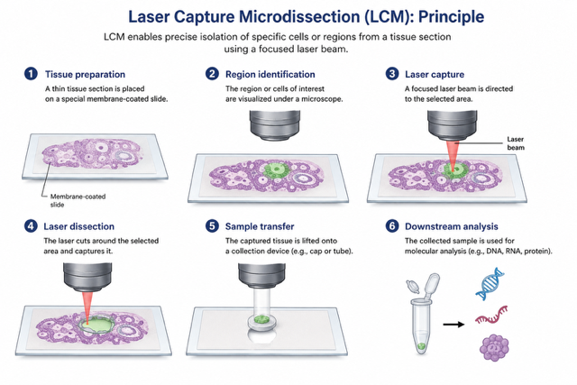
Laser Capture Microdissection (LCM), also known as Laser Microdissection (LMD), is a contact-free and contamination-free technique that enables the precise isolation of specific cells or tissue areas from a wide range of biological samples. This powerful method provides high accuracy and efficiency, making it ideal for genomic, proteomic, and cellular analysis.
VitroVivo’s LCM/LMD Services
VitroVivo Biotech offers advanced Laser Capture Microdissection services to support your research needs. By isolating individual cells or specific regions of tissue, we enable downstream analysis with unmatched precision for applications like next-generation sequencing, PCR, proteomics, and gene expression studies.
Laser Capture Microdissection (LCM/LMD) Applications
- Frozen & FFPE Sample LCM/LMD – Isolation from both frozen and formalin-fixed, paraffin-embedded (FFPE) tissue samples.
- Immunoguided LCM/LMD – Targeted isolation based on immunohistochemical markers.
- Live Cell Microdissection – Extraction of live cells for cloning, reculturing, or functional studies.
- Plant Cell LCM/LMD – Isolation of plant tissue cells for molecular research.
- Downstream Analysis – DNA, RNA, and protein extraction from dissected samples for genomic and proteomic analysis.
Advantages of LCM/LMD
✅ Contact-Free and Contamination-Free – Minimizes contamination risk and preserves sample integrity.
✅ High Precision – Laser cutting width of less than 1 µm ensures minimal disruption to surrounding tissue.
✅ Live Cell Viability – Isolate live cells without damaging them, enabling cloning and reculturing.
✅ Preserved Sample Quality – No alteration to the morphology or chemistry of isolated cells, making it ideal for DNA, RNA, and protein analysis.
Why Choose VitroVivo for LCM/LMD Services?
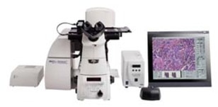
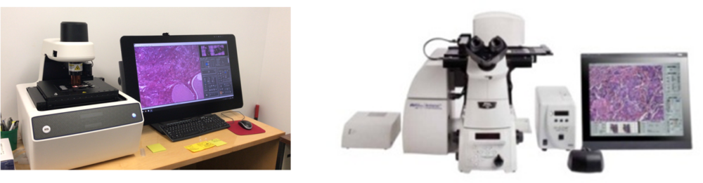
- State-of-the-Art Technology – Advanced laser capture microdissection techniques for accurate and reliable results.
- Experienced Professionals – Skilled technicians providing precise isolation for complex research needs.
- Custom Solutions – Tailored services for biomedical research, plant studies, and more.
📩 Contact VitroVivo Biotech today for Laser Capture Microdissection (LCM/LMD) services to elevate your research with high-quality cell isolation and analysis.
Why Choose VitroVivo for LCM/LMD Services? Watch the video below to learn more!
Frequently Asked Questions (FAQ)
FAQ 1. What is Laser Capture Microdissection (LCM) or Laser Microdissection (LMD) and its downstream applications ?
LCM or LMD is a method to isolate specific single cells or entire areas of tissue from a wide variety of tissue samples under direct microscopic visualization. LCM or LMD technology can harvest the cells of interest directly or can isolate specific cells by cutting away unwanted cells to generate histologically pure enriched cell populations. A variety of downstream applications exist: DNA genotyping and loss-of-heterozygosity (LOH) analysis, RNA transcript profiling, cDNA library generation, proteomics discovery and signal-pathway profiling.
FAQ 2. How do I prepare frozen sections for Laser Capture Microdissection (LCM/LMD)?
- General Guidelines for Specimen Preparation: Tissue should be cut and pieces frozen as soon as possible upon removal from the body or after death. RNase-Free conditions should be applied at all times during handling of tissues and sections.
- RNase-Free Technique: a). Wear disposable gloves and change frequently ; b). Use new or clean instruments between each animal or patient specimen; c). Use RNase-free or Nuclease free solutions, glassware and plasticware; d). Use RNase AWAY® or similar product to clean equipment
- Frozen Tissue Preparation: a). Tissues should be frozen in OCT or a similar product; b). Place a small amount of OCT on bottom of cryomold making sure there are no bubbles; c).Place tissue in the mold oriented such that the histologic region of interest is cut en face; d). Fill the mold with OCT to completely cover the mold making sure there are no bubbles (i.e. place OCT going from center to the periphery of the mold.
- Preferred Freezing Methods: a). Isopentane cooled over liquid Nitrogen; b). Isopentane cooled with dry ice; c). Other Freezing Methods: Dry Ice Alone (not optimal, slow) , Liquid Nitrogen (not optimal, sectioning difficulties) , Cryostat (NO!!!)
Note: Tissues can be sectioned immediately or stored in a –70C freezer - Frozen Tissue Sectioning: a).Gloves must be worn at all times; b).Cryostat must be cleaned prior to use. All surfaces must be wiped down with 95-100% ethanol, especially knife holder and anti-roll plate. c). Recommended Section thickness: LCM alone=8-10µm. d). Mount sections onto slides at room temperature. After mounting the sections should be frozen as quickly as possible by placing directly on dry ice. SLIDES MUST REMAIN COLD!!! e). For mounting of sections onto frame membrane slides refer to Arcturus Protocol; f). Use separate areas of the microtome blade for each specimen; g). Sections can be stored in a slide box in a –70ºC freezer until further processing.
- General Staining Guidelines: a). Total staining time should be as short as possible; b). Staining dishes should be nuclease free; c). Staining solutions should be dedicated for use with LCM samples; d). Solutions are prepared with nuclease free water; e). Stained slides can be held in xylene until initiation of laser capture microdissection; f). Once removed from xylene, microdissection should be completed within 2 hours
- Histochemical Staining: a) 75% ETOH – 30 secs; b) NF dH20 – 30 secs; c) H&E stain -10-30 secs; d) NF dH20 – 30 secs; e) 75% ETOH – 30 secs; f) 95% ETOH – 30 secs; g) 100% ETOH – 30 secs; h) Xylene – 5 mins.
FAQ 3. How do I prepare FFPE sections for Laser Capture Microdissection (LCM/LMD)?
- FFPE Tissue Preparation
Please Note: Formalin-fixation occurs by cross-linking proteins and nucleic acids with the aldehyde groups that not only affects the structural integrity of nucleic acids and proteins but the recovery as well. DNA can be analyzed most easily but there are greater challenges when analyzing RNA and proteins since the extraction process causes degradation of biomolecules.
a) Tissue should be placed in 10% Neutral Buffered Formalin as soon as possible after harvesting
b) Fixation should not exceed 24 hours at room temperature with tissue thickness not exceeding 5mm during fixation process
c) Tissues should undergo tissue processing with embedding in paraffin immediately after fixation. Storage in ethanol or PBS is not recommended - FFPE Tissue Sectioning: a) Use Nuclease Free or DEPC treated water for tissue floatation bath; b)Float sections for minimal amount of time, no more than 1-2 mins; c) Once sections mounted on slides, prop up vertically to allow water to drain away from sections; d) Air dry for about 2 hrs at room temperature. e) Do not use oven to dry sections; g). Slides can be store for up to 2 wks at room temperature with dessicant, for longer terms store at –70ºC.
- General Staining Guidelines: a) Total staining time should be as short as possible; b) Staining dishes should be nuclease free; c) Staining solutions should be dedicated for use with LCM samples; d) Solutions are prepared with nuclease free water; e) Stained slides can be held in xylene until initiation of laser capture microdissection; f) Once removed from xylene, microdissection should be completed within 2 hours.
- Histochemical Staining: a) Fresh xylenes (to depariffinize the sections) – 5 min; b) Fresh xylenes – 5 min; c) 100% ethanol – 15 sec; d) 95% ethanol – 15 sec; e) 70% ethanol – 15 sec; f) Deionized water – 15 sec; g) Mayer’s Hematoxylin – 30 sec; h) Deionized water – rinse (x 2) – 15 sec; i) 70% ethanol – 15 sec; j) Eosin Y – 5 sec; k) 95% ethanol – 15 sec; l) 95% ethanol – 15 sec; m) 100% ethanol – 15 sec; n) 100% ethanol – 15 sec; o) Xylenes (to ensure dehydration of the section) – 60 sec; p) Air-dry for approximately 2 minutes or gently use air gun to completely remove xylenes;
- The tissue is now ready for LCM process.
FAQ 4 Can you tell me the protocol for the preparation of rapid immunofluorence staining for direct laser capture of immunoreactive cells ?
- Outline tissue with a hydrophobic pen and allow to dry.
- Fix tissue in acetone-methanol (1:1) solution at -20 °C for 10 min. NOTE: Our experience has shown that the acetone-methanol fixation resulted in much more consistent immunohistochemistry than acetone or methanol alone.
- Rinse slide in phosphate buffered saline (PBS) with 1% Triton (RNase free).
- Cover sections with 100-200 µl PBS with 1% Triton with 1º antibody diluted optimized dilution with 400 U/ml RNasin. Incubate for 5-10 min.
- Rinse briefly in PBS twice and PBS-1% Triton.
- Cover tissue with 100-200 µl of goat anti-primary IgG labeled with Alexa Fluor 488 diluted 1:100 in PBS-1% Triton with 400 U/ml RNasin and 50 ng/ml DAPI. Incubate for 5 min.
- Rinse 2 times in PBS then dehydrate 30 s in a graded series of RNase free ethanol (75%-75%-95%-95%-100%-100%).
- Incubate in two washes of Xylene for 1 min then 5 min.
- Remove slides from Xylene immediately prior to use for LCM and allow to air dry.
FAQ 5. Does VitroVivo provide downstream application services of laser capture microdissection?
Yes, please send your request to: service@vitrovuvo.com. You also can visit the page of FFPE, frozen and laser microdissection sample analysis services.
FAQ 6. Can you show me some publications related laser capture microdissection in past 2 years?
Yes, see the links below:
- Genome-wide analysis revealed that DZNep reduces tubulointerstitial fibrosis via down-regulation of pro-fibrotic genes. Mimura I, et al.I.Sci Rep. 2018 Feb 28;8(1):3779.
- Alternative transcription of a shorter, non-anti-angiogenic thrombospondin-2 variant in cancer-associated blood vessels. Roudnicky F, et al. Oncogene. 2018 Feb 22.
- CrosstalkNet: A visualization tool for differential co-expression networks and communities. Manem VS, et al. Cancer Res. 2018 Feb 19.
- High-resolution spatiotemporal transcriptome mapping of tomato fruit development and ripening. Shinozaki Y, et al. Nat Commun. 2018 Jan 25;9(1):364.
- Lack of Fgf18 causes abnormal clustering of motor nerve terminals at the neuromuscular junction with reduced acetylcholine receptor clusters. Ito K, et al. Sci Rep. 2018 Jan 11;8(1):434
- Genomics-Driven Precision Medicine for Advanced Pancreatic Cancer: Early Results from the COMPASS Trial. Aung KL, et al. Clin Cancer Res. 2017 Dec 29.
- Morphological changes in different populations of bladder afferent neurons detected by herpes simplex virus (HSV) vectors with cell-type-specific promoters in mice with spinal cord injury. Shimizu N, et al. Neuroscience. 2017 Nov 19;364:190-201.
- Macrophage Infiltration Is a Causative Factor for Ligamentum Flavum Hypertrophy through the Activation of Collagen Production in Fibroblasts. Saito T, et al. Am J Pathol. 2017 Dec;187(12):2831-2840
- Defective decidualization during and after severe preeclampsia reveals a possible maternal contribution to the etiology.Garrido-Gomez T, et al. Proc Natl Acad Sci U S A. 2017 Oct 3;114(40):E8468-E8477.
- Cell-specific expression of plant nutrient transporter genes in orchid mycorrhizae. Fochi V, et al. Plant Sci. 2017 Oct;263:39-45. doi: 10.1016/j.plantsci.2017.06.015. Epub 2017 Jul 11.
- Heterogeneity of neuroblastoma cell identity defined by transcriptional circuitries. Boeva V, et al. Nat Genet. 2017 Sep;49(9):1408-1413.
- cGAS surveillance of micronuclei links genome instability to innate immunity. Mackenzie KJ, et al. Nature. 2017 Aug 24;548(7668):461-465. doi: 10.1038/nature23449. Epub 2017 Jul 24.
- The integrated pathway of TGFβ/Snail with TNFα/NFκB may facilitate the tumor-stroma interaction in the EMT process and colorectal cancer prognosis. Li H, et al.Sci Rep. 2017 Jul 7;7(1):4915.
- Interaction of reactive astrocytes with type I collagen induces astrocytic scar formation through the integrin-N-cadherin pathway after spinal cord injury. Hara M, et al. Nat Med. 2017 Jul;23(7):818-828.
- Increased T-cell Infiltration Elicited by Erk5 Deletion in a Pten-Deficient Mouse Model of Prostate Carcinogenesis. Loveridge CJ, et al. Cancer Res. 2017 Jun 15;77(12):3158-3168.
- Human Alternative Macrophages Populate Calcified Areas of Atherosclerotic Lesions and Display Impaired RANKL-Induced Osteoclastic Bone Resorption Activity.
- Chinetti-Gbaguidi G, et al. Circ Res. 2017 Jun 23;121(1):19-30.
- Maternal smoke exposure decreases mesenchymal proliferation and modulates Rho-GTPase-dependent actin cytoskeletal signaling in fetal lungs. Unachukwu U, et al. FASEB J. 2017 Jun;31(6):2340-2351.
- Synaptic Plasticity onto Dopamine Neurons Shapes Fear Learning. Pignatelli M, et al. Neuron. 2017 Jan 18;93(2):425-440.
- Laser microdissection of tomato fruit cell and tissue types for transcriptome profiling. Martin LB, et al.Nat Protoc. 2016 Dec;11(12):2376-2388.
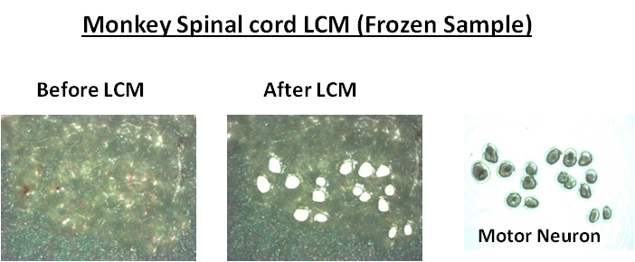
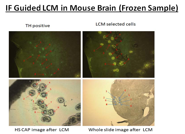
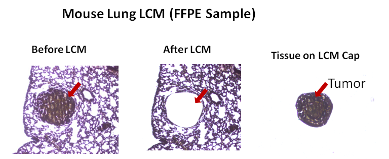
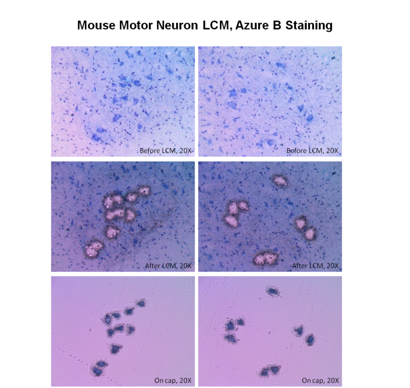
Example LCM Images