Description
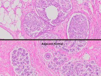
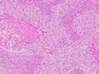
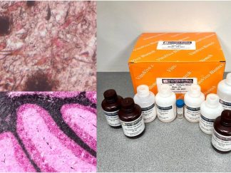
The VitroView™ Von Kossa Calcium Stain Kit is a reliable and easy-to-use solution for detecting calcium deposits in tissue samples. Designed for use in paraffin-embedded sections, undecalcified bone, and calcium-rich tissues, this kit provides rapid and reproducible staining, highlighting calcium salts in black or brown-black, with nuclei counterstained in red and cytoplasm in pink.
Ideal for histology, pathology, and biomedical research, the VitroView™ Von Kossa kit enables high-contrast visualization of calcium deposition, supporting studies of bone development, mineralization disorders, and tissue calcification. With optimized reagents and clear instructions, this kit delivers consistent, high-quality results for both routine and advanced research applications.
Key Benefits
- Sensitive and specific detection of calcium salts
- Compatible with paraffin sections and undecalcified tissues
- Quick and reproducible staining
- Optimized for both Sunlight and standard laboratory lighting
Kit Contents
| VB-3043-1 | Silver Nitrate Solution | 100 ml |
| VB-3043-2 | Sodium Thiosulfate Solution | 100 ml |
| VB-3043-3 | Nuclear Fast Red Solution | 100 ml |
Storage Condition:
Silver Nitrate Solution : Store at 2–8°C.
The others: Store at room temperature.
Staining Procedures for FFPE Tissue Sections
1. Deparaffinize in xylene I for 6 minutes and II for 6 minutes.
2. Rehydrate
1) Ethanol 100% (2 minutes)
2) Ethanol 100% (2 minutes)
3) Ethanol 95% (2 minutes)
4) Ethanol 95% (2 minutes)
3. Rinse sections in distilled water for 5 minutes.
4. Cover the sections with 0.5-1 ml of Silver Nitrate Solution and expose them to bright sunlight through a clear glass window for 20 minutes. Alternatively, place the sections under a 60–100 watt light bulb for 1 hour, or until the calcium turns black.
Note: If the stain is weak or fades, this indicates insufficient light exposure. In such cases, extend the incubation time as needed, up to several hours.
5. Rinse the sections thoroughly with multiple changes of distilled water.
6. To remove any unreacted silver, cover the sections with 0.5-1 ml of sodium thiosulfate and let it sit for 5 minutes.Rinse thoroughly in distilled water.
7. Counterstain with nuclear fast red for 5 minutes.
8. Rinse in distilled water.
9. Dehydrate with 2 changes of 95% Ethanol and 2 changes of 100% Ethanol (2 minute per change).
10. Clear with 3 changes of xylene (5 minute per change)
11. Mount coverslip onto glass slide with Permount or some other suitable organic mounting medium
Notes
1. UV light typically produces a stronger reaction, resulting in black calcium deposits. Exposure to a regular 60–100 watt light bulb often yields a brown-black coloration.
2. When confirmation is required, perform a negative control by treating a test slide with 10% formic acid for 10 minutes prior to Step 3. The treated slide should show no staining if the deposits are calcium.
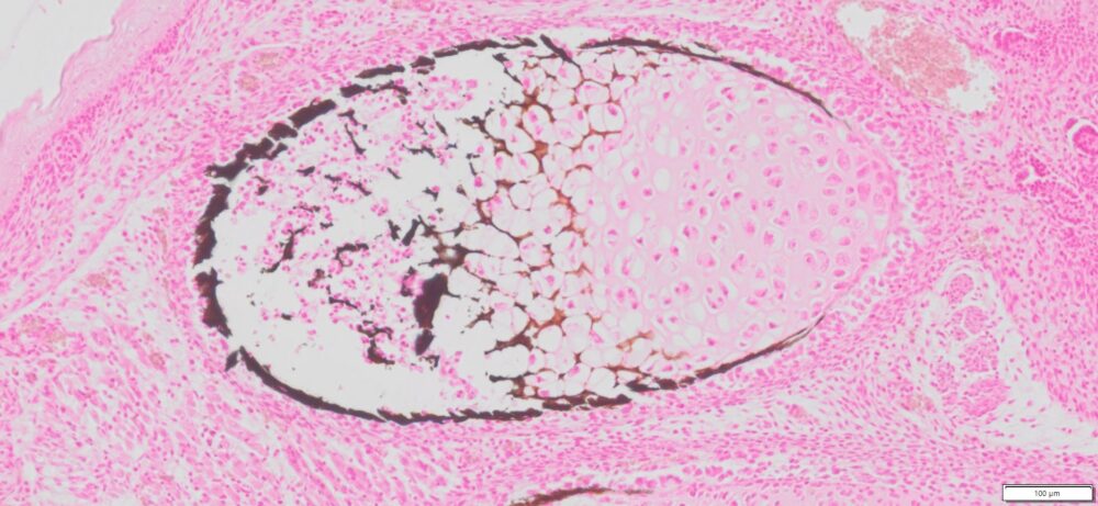
Expected Results
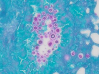
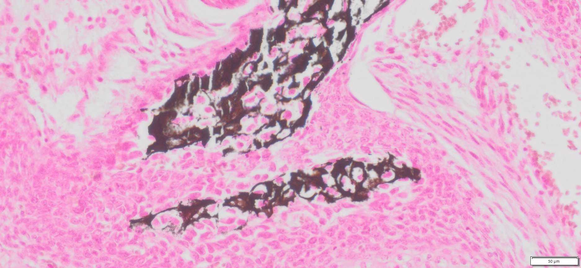
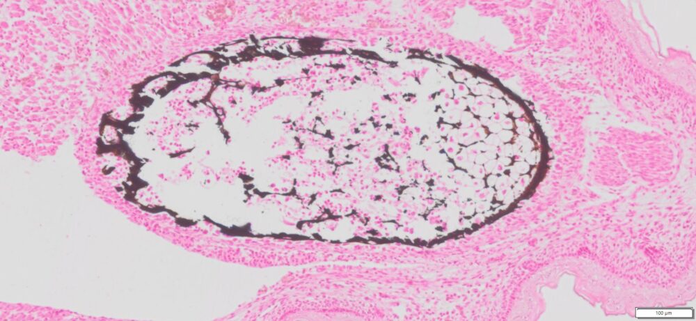
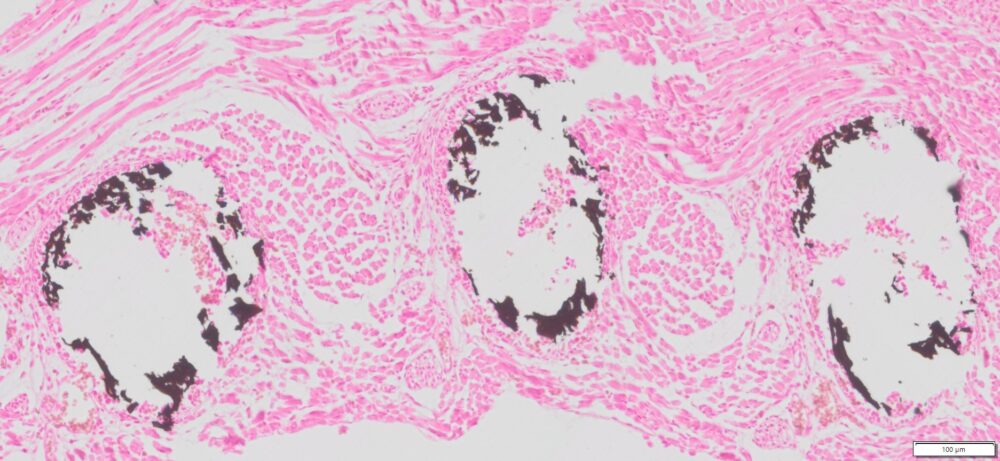
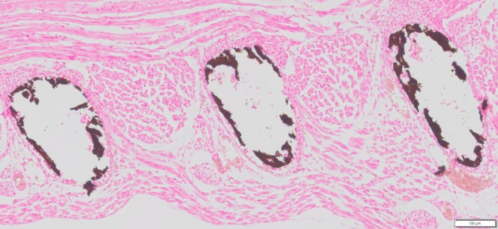
- Calcium salts———Black or brown-black
- Nuclei——————Red
- Cytoplasm————Pink
More Images
Precautions
- Handle reagents with care.
- Avoid contact with eyes, skin, or clothing.
- Do not ingest.
- Always wear gloves when handling chemicals.