Description
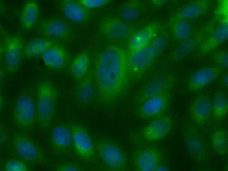
The plasma membrane also referred as cell membrane is a semipermeable lipid bilayer found in all cells that separates the interior of the cell from the exterior of the cell. It is responsible to regulate the transportation of materials and the movement of substances in and out of the cell. Here Vitrovivo Biotech offers a cell membrane fluorescence imaging probe that can identify cell types or cellular components in live and fixed cells.
DiIC18(5) is a far-red fluorescent (Ex:644nm/Em:665nm), lipophilic carbocyanine DiI analog with a longer-wavelength. It is weakly fluorescent in water but highly fluorescent and quite photostable when incorporated into membranes. It has an extremely high extinction coefficient and short excited-state lifetimes in lipid environments. Once applied to cells, the dye diffuses laterally within the plasma membrane.
Content
VB-1007 RTU Plasma Membrane Red Staining Solution——–30 ml
Storage Condition
Store at 2-8 °C and protect from light.
Application
Cell plasma membrane staining in both live and fixed cells.
General Protocol:
Adherent cells for fluorescence microscopy
- Grow cultured cells on sterile glass cover slips or slides overnight at 37 ºC.
- Follow appropriate protocol to fix cultured cells.
- Completely wash the cells with PBS as needed.
- Add adequate Read-to-Use Plasma Membrane Red Staining Solution to cover the whole sample.
- Incubate under dark at room temperature for 15-30 minutes.
- Rinse the sample several times with PBS and remove excess dye.
- Add antifade aqueous mounting medium and mount.
- Use appropriate filters and detect under fluorescence microscope according to standard protocol.
Suspension cells for fluorescence microscopy
- The cells are harvested into a 15 mL polypropylene centrifuge tube and spin down for 8 min at 600 RPM,
- The supernatant is discarded and the cells are resuspended in 0.5 ml of culture medium
- 1-2 drops of the cell suspension were placed on a slide in the central area and moved around to form a thin and even film with a glass spreader.
- (Option) You also can use cytocentrifuge to prepare cell slides.
- Air dry and follow appropriate protocol to fix cultured cells.
- Drop adequate RTU DAPI Nuclear Staining Solution to cover the whole sample on slide.
- Incubate under dark at room temperature for 15-30 minutes.
- Cover with coverslip and view under fluorescence microscope according to standard protocol.
Note: This product is intended for research purposes only. This product is not intended to be used for therapeutic or diagnostic purposes in humans or animals.
References
- Bohland JW, et al. A Proposal for a Coordinated Effort for the Determination of Brainwide Neuroanatomical Connectivity in Model Organisms at a Mesoscopic Scale. PLoS Computational Biology (2009) 5: e1000334
- Kuschel C, et al. Cell adhesion profiling using extracellular matrix protein microarrays. BioTechniques (2006) 40: 523-531
- Bianchi L, et al. A Potassium Channel-MiRP Complex Controls Neurosensory Function in Caenorhabditis elegans. THE JOURNAL OF BIOLOGICAL CHEMISTRY (2003) 278: 12415–12424