Description
The plasma membrane also referred as cell membrane is a semipermeable lipid bilayer found in all cells that separates the interior of the cell from the exterior of the cell. It is responsible to regulate the transportation of materials and the movement of substances in and out of the cell. Here VitroVivo Biotech offers a cell membrane fluorescence imaging probe that can identify cell types or cellular components in live and fixed cells.
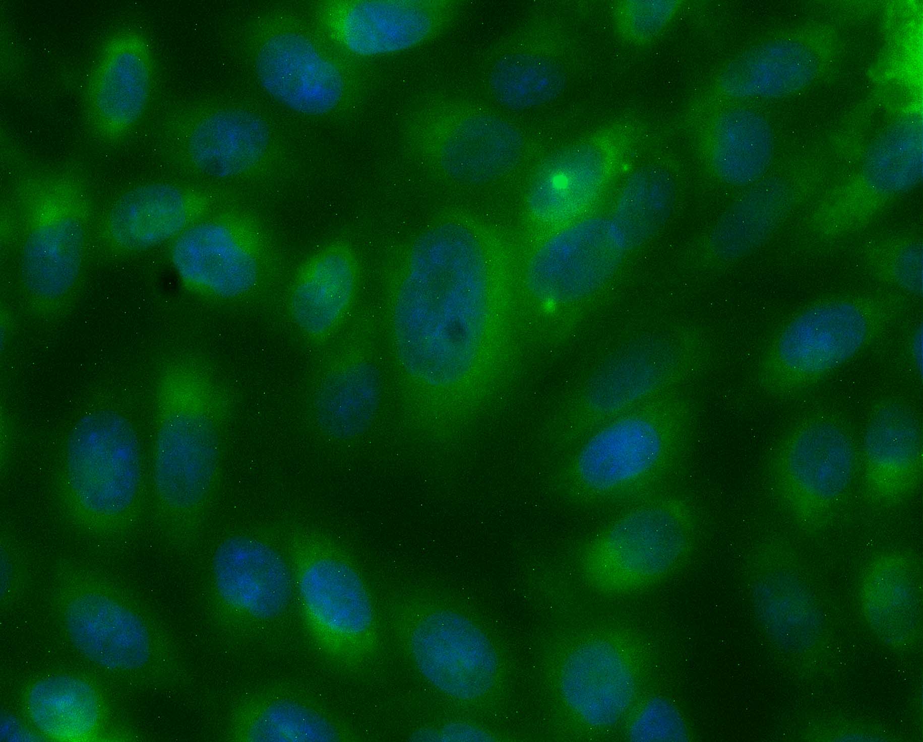
DiOC18(3) is a green fluorescent (Ex:485nm/Em:501nm), lipophilic carbocyanine dye that is widely used as a lipophilic tracer. It is weakly fluorescent in water but highly fluorescent and quite photostable when incorporated into membranes. It has an extremely high extinction coefficient and short excited-state lifetime in lipid environments. Once applied to cells, the dye diffuses laterally within the plasma membrane.
Content
VB-1006 RTU Plasma Membrane Green Staining Solution——–30 ml
Storage Condition
Store at 2-8 °C and protect from light.
Application
Cell plasma membrane staining in both live and fixed cells.
General Protocol:
Adherent cells for fluorescence microscopy
- Grow cultured cells on sterile glass cover slips or slides overnight at 37 ºC.
- Follow appropriate protocol to fix cultured cells.
- Completely wash the cells with PBS as needed.
- Add adequate RTU Plasma Membrane Green Staining Solution to cover the whole sample.
- Incubate under dark at room temperature for 15-30 minutes.
- Rinse the sample several times with PBS and remove excess dye.
- Add antifade aqueous mounting medium and mount.
- Use appropriate filters and detect under fluorescence microscope according to standard protocol.
Suspension cells for fluorescence microscopy
- The cells are harvested into a 15 mL polypropylene centrifuge tube and spin down for 8 min at 600 RPM,
- The supernatant is discarded and the cells are resuspended in 0.5 ml of culture medium
- 1-2 drops of the cell suspension were placed on a slide in the central area and moved around to form a thin and even film with a glass spreader.
- (Option) You also can use cytocentrifuge to prepare cell slides.
- Air dry and follow appropriate protocol to fix cultured cells.
- Drop adequate RTU Plasma Membrane Green Staining Solution to cover the whole sample on slide.
- Incubate under dark at room temperature for 15–30 minutes.
- Cover with coverslip and view under fluorescence microscope according to standard protocol.
Note: This product is intended for research purposes only. This product is not intended to be used for therapeutic or diagnostic purposes in humans or animals.
References
- Obregon C, et al. Exovesicles from Human Activated Dendritic Cells Fuse with Resting Dendritic Cells, Allowing Them to Present Alloantigens. The American Journal of Pathology (2006) 169: 2127-2136
- Boullier A, et al. The Binding of Oxidized Low Density Lipoprotein to Mouse CD36 Is Mediated in Part by Oxidized Phospholipids That Are Associated with Both the Lipid and Protein Moieties of the Lipoprotein. THE JOURNAL OF BIOLOGICAL CHEMISTRY (2000) 275: 9163–9169
- Gaub H, et al. Lateral Diffusion and Phase Separation in Two-Dimensional Solution of Polymerized Butadiene Lipid in Dimyristoylphosphatidylcholine Bilayers. BioPhys. J. (1984) 45:725-731
User Manual and Material Safety Data Sheet (MSDS) (PDF )