Description
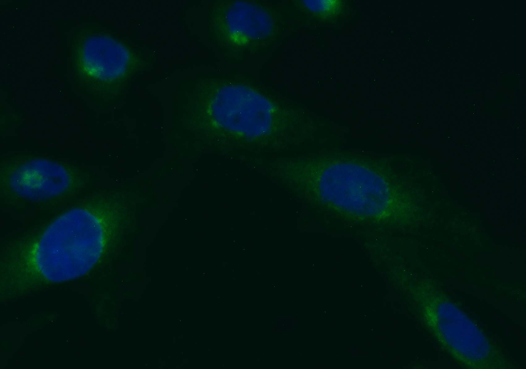
The Golgi apparatus is a folded flattened membrane-enclosed organelle that consists of multiple compartments within the cytoplasm of most eukaryotic cells, whose function is involved in protein secretion and intracellular transport of lipids and carbohydrates released from the endoplasmic reticulum (ER) to export outside of the cell. It also resides at the intersection of the secretory, lysosomal, and endocytic pathways. Here Vitrovivo Biotech offers a cell-permeant probe that can be used to distinguish the Golgi morphology in both live and fixed cells. Additionally, this product has application in lipid metabolism, trafficking studies.
NBD C6-ceramide is a sensitive fluorescent dye (Ex:466nm/Em:536nm) working in aprotic solvents and other nonpolar environments. It can be used as a selective stain for the Golgi apparatus in live and fixed cells. It is also used to study sphingolipid transport and metabolism mechanisms.
Content
VB-1005 RTU Golgi Staining Solution——–30 ml
Storage Condition
Store at 2-8 °C and protect from light.
Application
Cell Golgi staining in both live and fixed cells.
General Protocol:
Adherent cells for fluorescence microscopy
- Grow cultured cells on sterile glass cover slips or slides overnight at 37 ºC.
- Follow appropriate protocol to fix cultured cells.
- Completely wash the cells with PBS as needed.
- Add adequate RTU Golgi Staining Solution to cover the whole sample.
- Incubate under dark at room temperature for 15-30 minutes.
- Rinse the sample several times with PBS and remove excess dye.
- Add antifade aqueous mounting medium and mount.
- Use appropriate filters and detect under fluorescence microscope according to standard protocol.
Suspension cells for fluorescence microscopy
- The cells are harvested into a 15 mL polypropylene centrifuge tube and spin down for 8 min at 600 RPM,
- The supernatant is discarded and the cells are resuspended in 0.5 ml of culture medium
- 1-2 drops of the cell suspension were placed on a slide in the central area and moved around to form a thin and even film with a glass spreader.
- (Option) You also can use cytocentrifuge to prepare cell slides.
- Air dry and follow appropriate protocol to fix cultured cells.
- Drop adequate RTU Golgi Staining Solution to cover the whole sample on slide.
- Incubate under dark at room temperature for 15-30 minutes.
- Cover with coverslip and view under fluorescence microscope according to standard protocol.
Note: This product is intended for research purposes only. This product is not intended to be used for therapeutic or diagnostic purposes in humans or animals.
References
- Scidmore MA, et al. Restricted Fusion of Chlamydia trachomatis Vesicles with Endocytic Compartments during the Initial Stages of Infection. INFECTION AND IMMUNITY (2003) 71: 973–984
- Miura I, et al. (Effects of Mepanipyim on Intracellular Trafficking: A Comparative Study on Its Effects on Exocytic and Endocytic Trafficking of Proteins, Sphingolipids, and Cholesterol. Biosci. Biotech. Biochem. (1996) 60: 1690-1697
- Pagano RE, et al. Molecular Trapping of a Fluorescent Ceramide Analogue at the Golgi Apparatus of Fixed Cells: Interaction with Endogenous Lipids Provides a trans-Golgi Marker for Both Light and Electron Microscopy. The Journal of Cell Biology (1989) 109: 2067-2079