Description
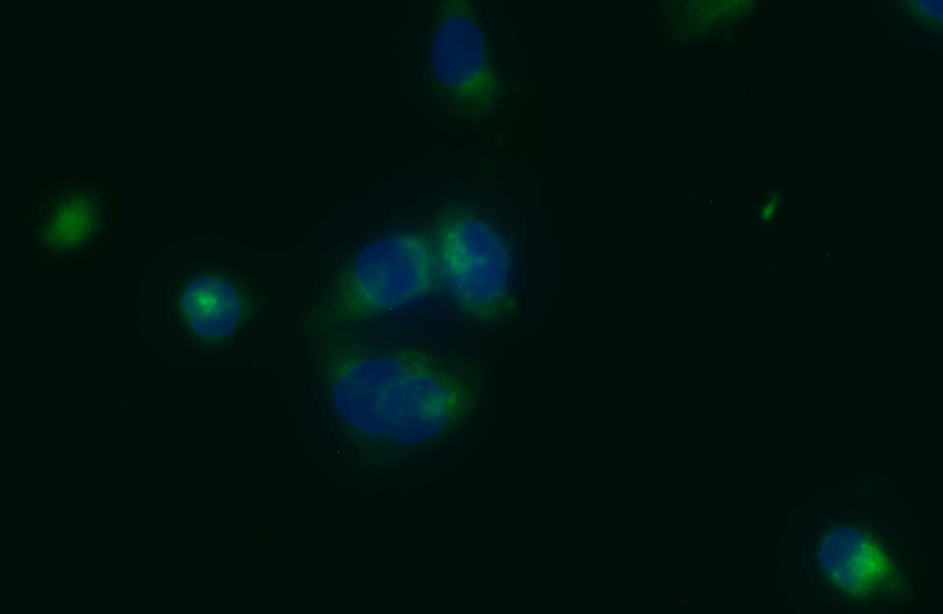
Mitochondrion is a double-membrane organelle found in most eukaryotic cells, whose key function is energy production through oxidative phosphorylation and lipid oxidation. The mitochondria consist of outer membrane and inner membrane extending into the inner matrix with various structures depending on cell type and intracellular metabolic state. Mitochondria are also involved in other metabolic functions. Here VitroVivo Biotech offers a fluorescent dye used in fluorescence microscopy, flow cytometry and fluorescence correlation spectroscopy studies in living cells.
Rhodamine123 is a cationic cell-permeant, green-fluorescent dye (Ex:507nm/Em:529nm) that is taken by active mitochondria without cytotoxic effects. This dye stains mitochondria in living cells depending on membrane potential and does not stain Endoplasmic Reticulum (ER).
Content
VB-1003 RTU Mitochondrion Staining Solution——–30 ml
Storage Condition
Store at 2-8 °C and protect from light.
Application
Cell nuclear staining in living cells
General Protocol:
Adherent cells for fluorescence microscopy
- Grow cultured cells on sterile glass cover slips or slides overnight at 37 ºC.
- Follow appropriate protocol to fix cultured cells.
- Completely wash the cells with PBS as needed.
- Add adequate RTU Mitochondrion Staining Solution to cover the whole sample.
- Incubate under dark at room temperature for 15-30 minutes.
- Rinse the sample several times with PBS and remove excess dye.
- Add antifade aqueous mounting medium and mount.
- Use appropriate filters and detect under fluorescence microscope according to standard protocol.
Suspension cells for fluorescence microscopy
- The cells are harvested into a 15 mL polypropylene centrifuge tube and spin down for 8 min at 600 RPM,
- The supernatant is discarded and the cells are resuspended in 0.5 ml of culture medium
- 1-2 drops of the cell suspension were placed on a slide in the central area and moved around to form a thin and even film with a glass spreader.
- (Option) You also can use cytocentrifuge to prepare cell slides.
- Air dry and follow appropriate protocol to fix cultured cells.
- Drop adequate RTU Mitochondrion Staining Solution to cover the whole sample on slide.
- Incubate under dark at room temperature for 15–30 minutes.
- Cover with coverslip and view under fluorescence microscope according to standard protocol.
Note: This product is intended for research purposes only. This product is not intended to be used for therapeutic or diagnostic purposes in humans or animals.
References
- Ali H, et al. Furan-Conjugated Tripeptides as Potent Antitumor Drugs. Biomolecules (2020) 10:1684
- Follstad B D, et al. Mitochondrial membrane potential differentiates cells resistant to apoptosis in hybridoma cultures. Eur. J. Biochem. (2000) 267: 6534-6540
- Grimm D, et al. Establishment and characterization of a human papillary thyroid carcinoma cell line with oxyphilic differentiation (ONCO-DG 1). Virchows Archiv B Cell Pathol (1992) 62:97-104