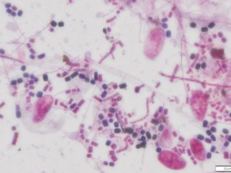
Description
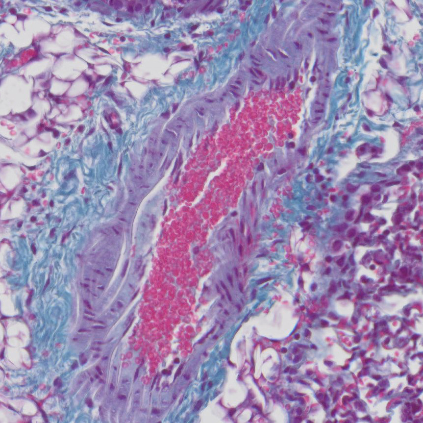
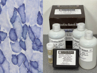
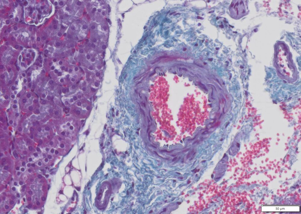
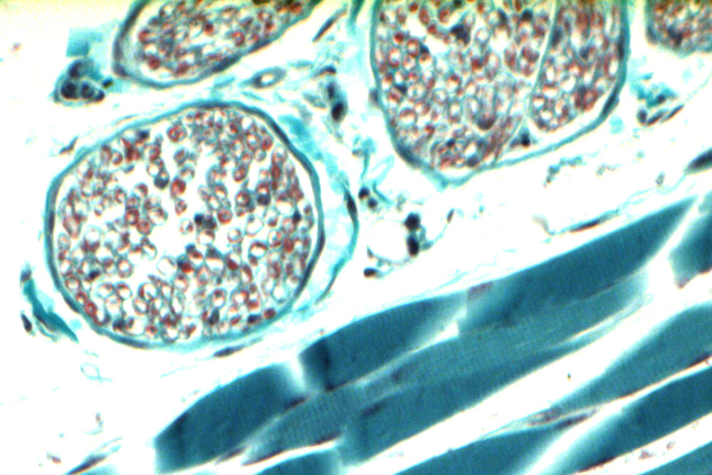
Trichrome stain is one of the most highly utilized special stains in the histopathology laboratory. Most of common uses for trichrome staining are liver biopsies, renal biopsies, dermatopathology, cardiac biopsies and muscle and nerve biopsies. Gomori’s trichrome is a one-step staining procedure that combines plasma stain (chromotrope 2R) and connective fiber stain (fast green FCF) in a phosphotungstic acid solution to which glacial acetic acid has been added. This kit is designed for both formalin-fixed and paraffin-embedded (FFPE) tissue and f rozen tissue section.
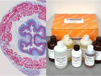
Kit Contents
- VB-3014-1 Gomori’s Trichrome solution ————250 ml
- VB-3014-2 Harris Hematoxylin solution————-250 ml
Storage Condition
Room temperature.
Protocol
- Sample preparation: 1) Frozen tissue: Cut 10 – 16 micron (12 µm) sections in a cryostat from snap frozen tissue. Fixation is not needed. 2) FFPE section: Cut 4-5 µm sections, deparaffinize in xylene for 6 min×2, rehydrate in ethanol 100% (2 min×2), ethanol 95% (2 min×2), and ethanol 70% (2 min×2), rinse in distilled water (5 minutes ×1).
- Immerse sections in Harris Hematoxylin solution for 5 minutes.
- Wash with tap water until the water is clear.
- Immerse sections in Gomori trichrome stain solution for 10 minutes.
- Differentiate using 0.2% acetic acid. A few dips should be sufficient.
- Immerse rack with sections directly into 95 % alcohol
- Continue to dehydrate in ascending alcohol solutions (95%× 2, 100%×2).
- Clear with xylene (5 min×2).
- Mount coverslip onto the slide with Permount or some other suitable organic mounting medium.
Expected Results
- Nuclei: dark blue
- Muscle myofibrils: green-blue
- Mitochondria and endoplasmic reticulum stain: red
- Connective tissue stains: pale green-blue
- Myelin stains: purple red
- Type 1 fibers stain darker blue/green as compared to type 2 fibers.
More Imnages
Precautions
Handle with care. Avoid contact with eyes, skin and clothing. Do not ingest. Wear gloves

