Description
Sample Basic Information
| Sample ID | Organ | Pathology Diagnosis | Gender | Age | Grade | TMN | IHC Data | Sample Format |
| 02001T | Human breast | Human breast invasive ductal carcinoma | Female | 45 | 2 | N/A | ER(80% +), PR(70%+), E-cad(+), P120(+), CK5/6(-), CK8/18(+), Ki67(15% +), Her2 (-) | FFPE |
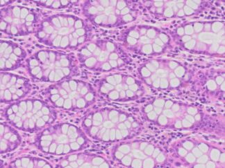
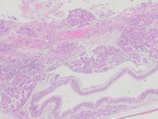
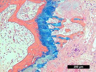
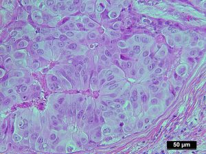
Tissue H&E Staining and IHC images
H&E Stain |
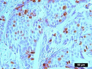
Ki 67 IHC |
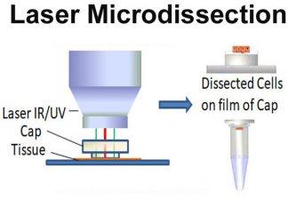
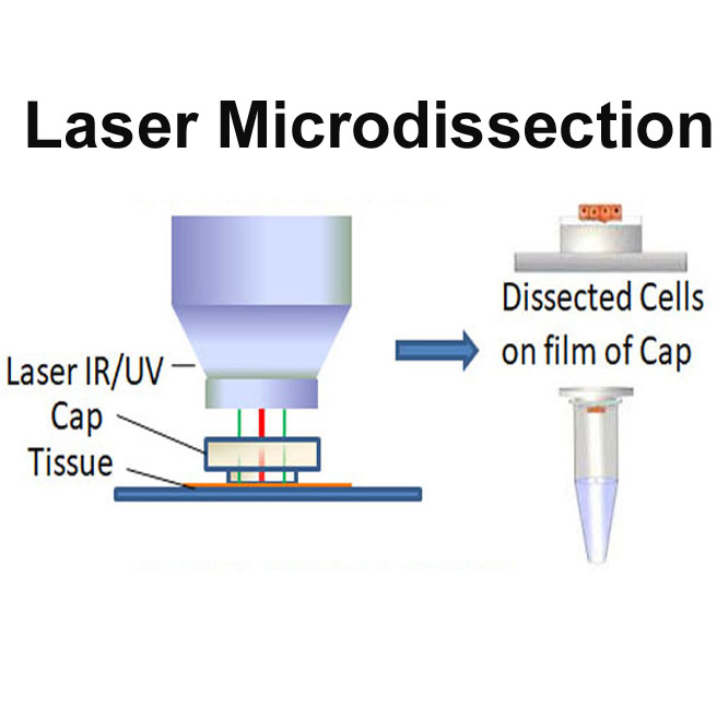
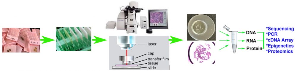
Flowchart of LCM for The Product Preparation
- FFPE samples are cut at 6 microns and mounted onto pen membrane glass slides and let the slides dry for 2 hours at room temperature.
- After deparaffinization and rehydration, quick H&E staining is performed.
- Arcturus XT Laser Microdissection System Tissue is used for LCM. Microdissection is performed using IR and UV laser and CapSure Macro LCM Cap.
- The microdissected cells are on the film of Macro LCM Cap. The total dissected cells may be varied between 10,000 to 50,000 cells from different tissue type.
- Extract protein from LCM Cap.
Quality Control
- The tissue H&E staining slides were examined by certified pathologists. Pathological re-confirmation report is generated and digital image captured.
- LCM is supervised by our certified pathologist.
- Three example LCM images including Before, After LCM, and LCM cap images from LCM tissue slides are took to demonstrate the dissected cells.
Extraction Protocol for Protein from LCM Caps
- For protein extraction, you can follow the protocol from Longuespée R, et al. A laser microdissection-based workflow for FFPE tissue microproteomics: Important considerations for small sample processing, Methods;2016, 104: 154-162
Product Format and Size:
Protein in 50 μl of lysis buffer from 2 LCM caps.
Applications
- Protein from pure cell population microdessected from FFPE tissues can be used for proteomics.
- The product is only used for biomedical research. It is not used for clinical diagnosis.
Storage
Store at -20ºC for shot time and at -80ºC for longer time
References
- Longuespée R, et al. A laser microdissection-based workflow for FFPE tissue microproteomics: Important considerations for small sample processing, Methods;2016, 104: 154-162
- Cai J, et al. The Use of Laser Microdissection in the Identification of Suitable Reference Genes for Normalization of Quantitative Real-Time PCR in Human FFPE Epithelial Ovarian Tissue Samples. PLoS ONE 2014, 9(4): e95974.
- Drummond ES, et al. Proteomic analysis of neurons microdissected from formalin-fixed, paraffin-embedded Alzheimer’s disease brain tissue.Sci Rep. 2015;5:15456
- Bockmeyer CL, et al. Recommendations for mRNA analysis of micro-dissected glomerular tufts from paraffin-embedded human kidney biopsy samples. BMC Mol Biol. 2018;19(1):2
- Murray, Graeme I. (Ed.). Laser Capture Microdissection (Methods and Protocols, © 2018