Description
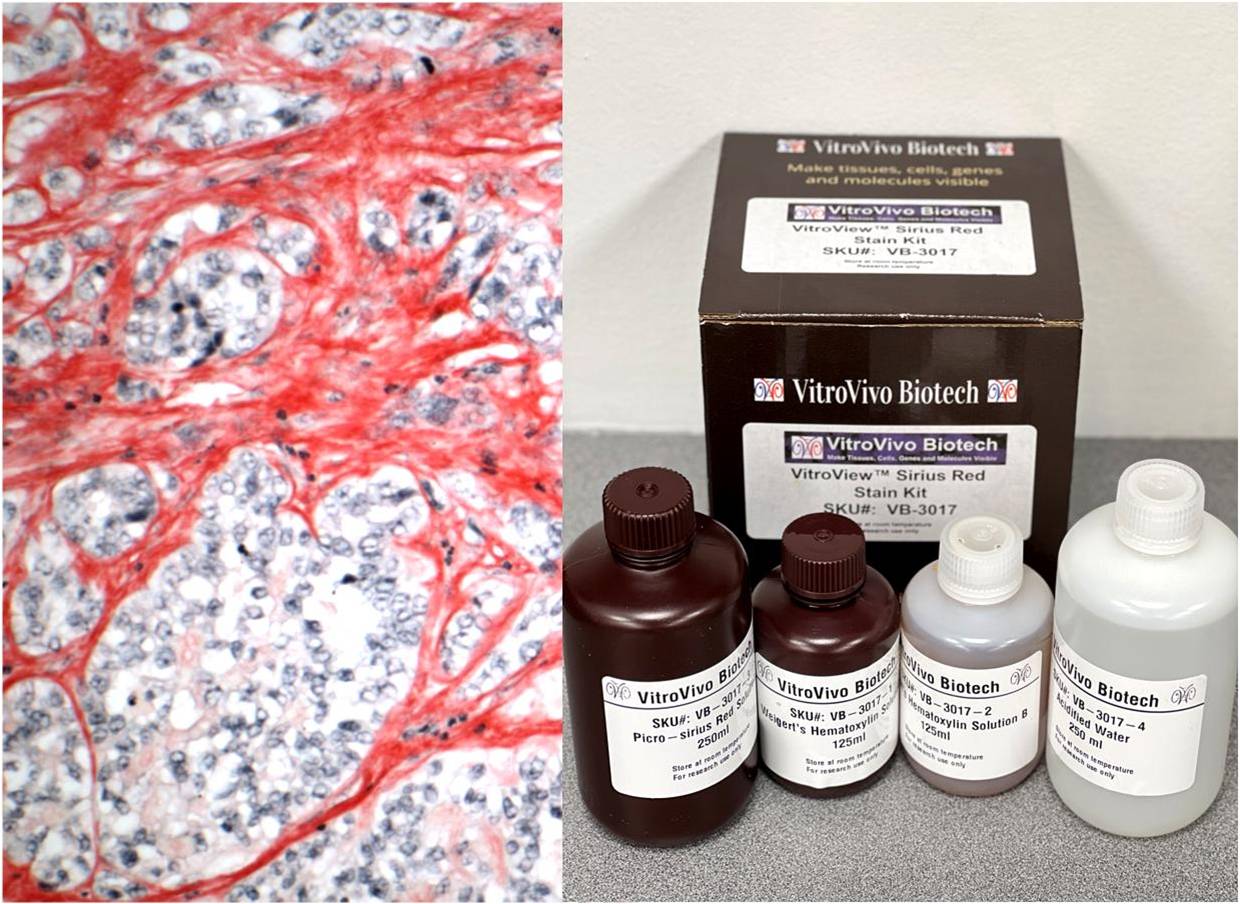
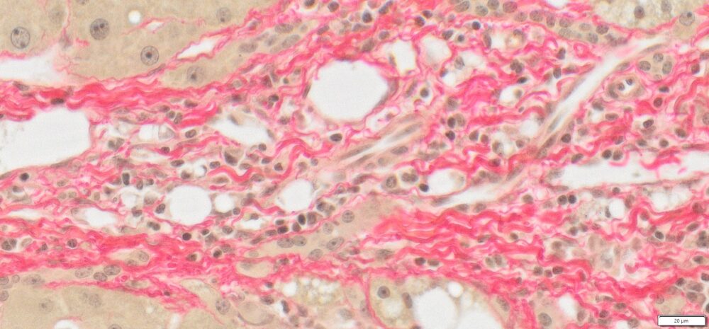
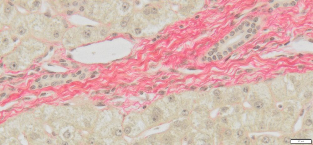
Specific staining by Picro-sirius red is one of the most important stains to study collagen networks in different tissues. VitroView™ Picro-Sirius Red Stain Kit is designed for stain of collagen I and III fibers. The Sirius Red stain may be viewed using standard light microscopy or polarized light resulting in birefringence of the collagen fibers to distinguish between type I and type III.
VitroView™ Picro-Sirius Red Stain Kit Introduction Video

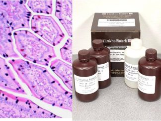
Kit Components
- VB-3017-1 Weigert’s Hematoxylin Solution A——–125 ml
- VB-3017-2 Weigert’s Hematoxylin Solution B ——-125 ml
- VB-3017-3 Picro-sirius Red Solution———————-250 ml
- VB-3017-4 Acidified Watetr————————————250 ml
Storage Condition
Room temperature.
Protocol
- a).For FFPE slides: Deparaffinize in Xylene I for 6 minutes and II for 6 minutes. Rehydrate: ethanol 100% (2 minutes)×2; ethanol 95% (2 minutes)×2; ethanol 70% (2 minutes)×1; b). For Frozen Sections: fix frozen sections in 10% formalin for 30 minutes.
- Rinses in distilled water 2min×3.
- Prepare Weigert’s working hematoxylin: mix Weigert’s Hematoxylin Solution A and B at 1:1 ratio.
- Stain nuclei with Weigert’s haematoxylin for 8 minutes, and then wash the slides for 10 minutes in running tap water.
- Stain in Picro-sirius Red Solution for 1 hour. Note: This gives near-equilibrium staining, which does not increase with longer times. Shorter times should not be used, even if the colors look good.
- Wash in two changes of acidified water.
- Wash slides, 2min × 2 in dH2O.
- Dehydrate with 2 changes of 95% Ethanol and 2 changes of 100% Ethanol (2 minute per change).
- Clear with 3 changes of xylene (5 minute per change) and coverslip with Permount or other suitable organic mounting medium.
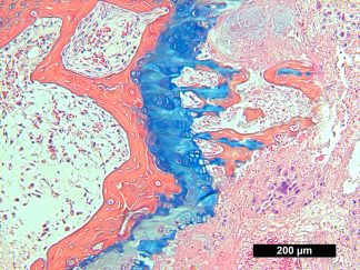
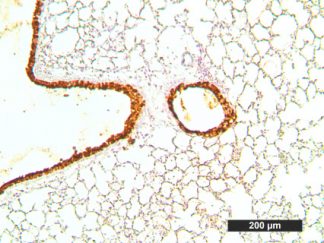
Expected results
Light Microscopy
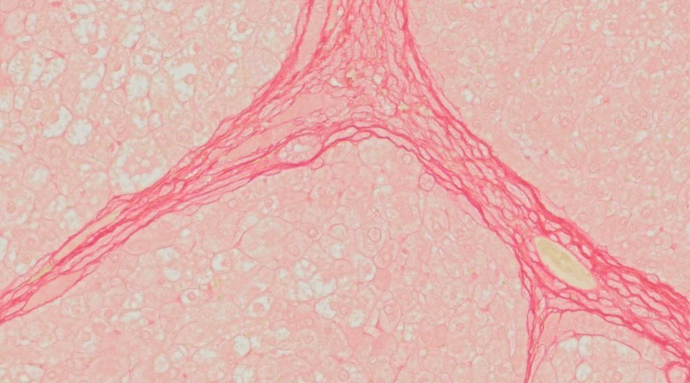
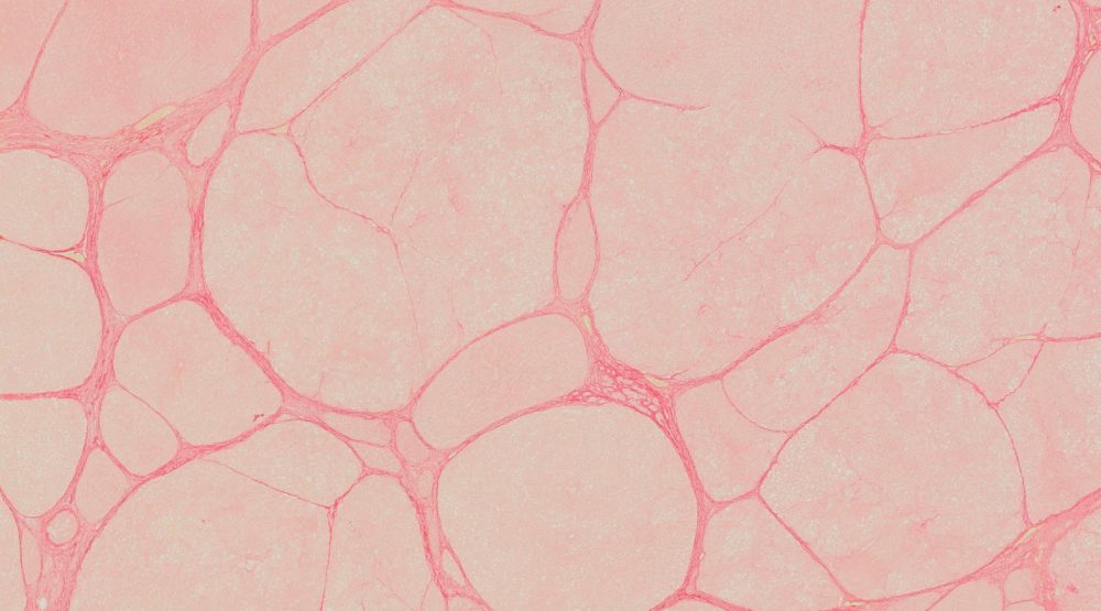
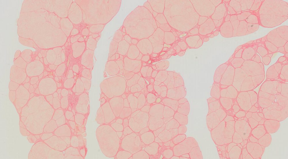
- Collagen ————————————————————– red
- Muscle Fibers —————————————————– yellow
- Cytoplasm ———————————————————- yellow
Polarized Light Microscopy
- Type I (Thick fibers) —————————————- yellow‐orange
- birefringence Type III (Thin fibers) ——————- green birefringence
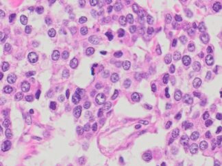
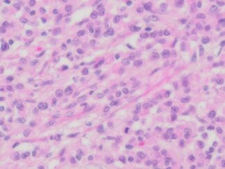
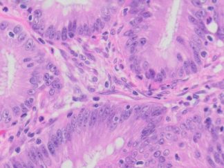
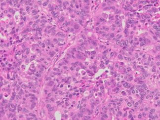
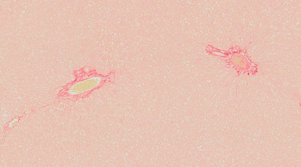
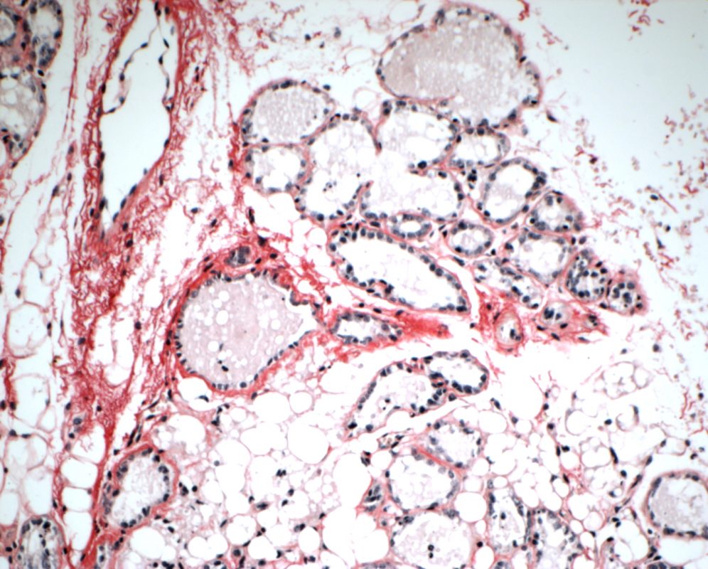
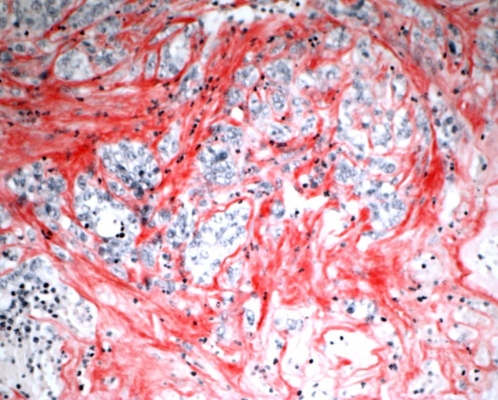
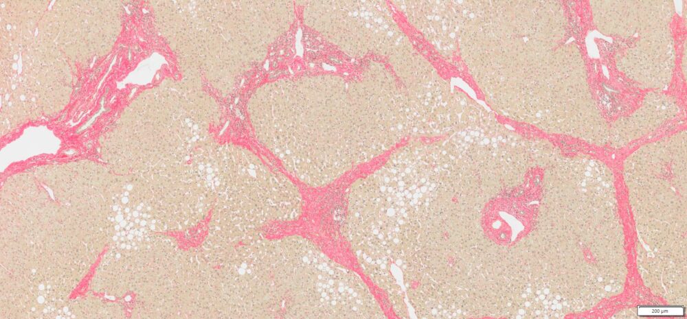
More Pico-Sirius Red Staining Images
|
|
|
|
|
|
|
|
|
|
|
|
|
|
Note: VitroView™ Picro-Sirius Red Stain Kit is intended for research purposes only. This product is not intended to be used for therapeutic or diagnostic purposes in humans or animals.
Precautions: Handle with care. Avoid contact with eyes, skin and clothing. Do not ingest. Wear gloves.
Peer-reviewed publications and preprints that have cited the use of VitroView™ Picro-Sirius Red Stain Kit (VB-3017)
-
Kin J (2023)-TLR7 activation by miR-21 promotes renal fibrosis by activating the pro-inflammatory signaling pathway in tubule epithelial cells
Cell Communication and Signaling (2023) 21:215 — This study used VitroView™ Picro-Sirius Red Stain Kit (VB-3017) to visualize collagen I and III in kidney sections to assess the degree of renal fibrosis. Springer Nature Link - Kim YY, et al (2022) — Genetic Deletion of Syndecan-4 Alters Body Composition, Metabolic Responses, and Collagen Deposition in High-Fat Diet-Induced Obesity.
Nutrients 11(11):2810. Staining of Type I and III collagen fibers was performed with a Picro-Sirius Red staining kit (Cat. No. VB-3017, VitroVivo Biotech, Rockville, MD, USA) to assess collagen deposition in adipose tissues. MDPI -
Ha S, et al (2022)-Activation of PAR2 promotes high-fat diet-induced renal injury by inducing oxidative stress and inflammation
International Journal of Molecular Sciences / ScienceDirect 1868 (10), 166474— Sirius Red staining was performed using a commercially available kit from VitroVivo (VB-3017) to determine renal fibrosis and damage. ScienceDirect -
Marini V, et al (2022)-Long-term culture of patient-derived cardiac organoids recapitulated Duchenne muscular dystrophy cardiomyopathy and disease progression
PMC (National Institutes of Health) 10.3389/fcell.2022.878311 — This article describes using the VitroView™ Picro-Sirius Red Stain Kit (VB-3017) to evaluate collagen deposition in cardio-related organoid tissue (used according to manufacturer’s instructions). PMC -
Giarratana et al (2020)-MICAL2 is essential for myogenic lineage commitment (PMC) Cell Death and Disease (2020) 11:654 — Picro-Sirius Red staining was carried out using Vitro View™ Picro-Sirius Red Stain Kit (VB-3017) in mouse tissue sections to dye connective tissue collagen. PMC
-
Lu A, et al (2025)-Muscle-specific ERRγ activation mitigates muscle atrophy after ACL injury FASEB Journal 39 (4): e70409 — This research reports H&E and Picro-Sirius Red stain (VitroView™ VB-3017, VitroVivo Biotech) staining following the manufacturer’s protocol for fibrosis/collagen visualization in muscle tissue. FASEB Journal