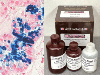
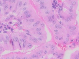
Description
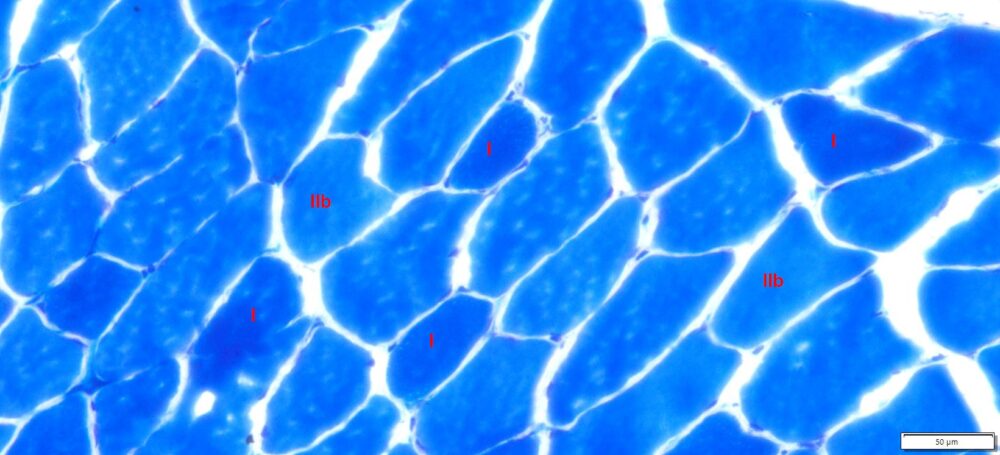
The metachromatic myofibrillar ATPase stain is a histochemical staining method used in the field of histology to identify different types of muscle fibers based on their ATPase activity. This stain helps differentiate muscle fibers into different types, primarily Type I (slow-twitch) and Type II (fast-twitch) muscle fibers.
VitroView™ Metachromatic Myofibrillar ATPase Stain Kit is used for studies related to muscle physiology, fiber typing, and the characterization of muscle fiber types in various tissues. It provides information about the distribution and characteristics of muscle fiber types based on their metabolic properties.
Kit Components
| SKU# | Reagent | Size |
| VB-3030-1 | Pre-Incubation Solution | 100ml |
| VB-3030-2 | Wash Buffer | 250ml |
| VB-3030-3 | ATP Incubation Solution | 1.5 ml ×5 |
| VB-3030-4 | 1 % Calcium Chloride Solution | 250ml |
| VB-3030-5 | Toluidine Blue Solution | 100 ml |
Storage
Store the ATP Incubation Solution at -20°C, and keep the other solution at room temperature.
Protocol
Tissue preparation for cryosectioning
- Sacrifice the animal using either cervical dislocation or decapitation, following ethical permit guidelines.
- Quickly collect tissues of interest without fixation, and promptly freeze them on dry ice (tissues may require freezing in isopentane) or propane chilled with liquid nitrogen to achieve optimal morphology.
- Store tissues in aluminum foil at -80°C until ready to section.
- Skeletal muscle tissue obtained was oriented for cross-sectioning in O.C.T. compound.
- Collect cryostat sections with a thickness ranging from 5-15μm (typically 10 μm). Thaw the sections onto slides at room temperature for 2-5 minutes, and store the slides without cover-slipping at -80°C until they are ready to be used.
Staining Procedure
- Apply 80~200μl of Pre-Incubation Solution onto dried frozen sections for 2 minutes, or place slides with dried frozen sections in the pre-incubation solution for 2 minutes at room temperature.
Note: Dropping the solution or buffer onto slides can conserve reagents and enhance slide staining efficiency. If immersing slides in the staining jar, a larger amount of reagents may be required.
- Drop 3-5 drops of Wash Buffer onto tissue sections, repeating three times, with a 2-minute interval for each application. Alternatively, immerse slides in a staining jar through three rinses in Wash Buffer, allocating 2 minutes for each rinse.
- Immediately cover the tissue section on the slides with 80~200μl of ATP Incubation Solution in humidity chamber and incubate for 25 minutes at 37°C.
Note: The ATP Incubation Solution may appear cloudy, but it can still be used.
- Rinse slides three times in 1% calcium chloride.
- Stain slides in toluidine blue solution for 1 minute.
- Rinse slides (maximum 30 seconds) in distilled water to remove excess stain from the slides and sections.
- Dehydrate rapidly: five dips each in one change of 95% ethanol and two changes of absolute ethanol.
- Clear the slides in two changes of xylene, each for 5 minutes, and mount with Permount.
Positive Control
Snap frozen striated muscle
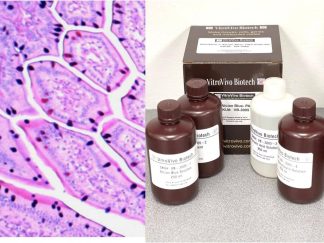
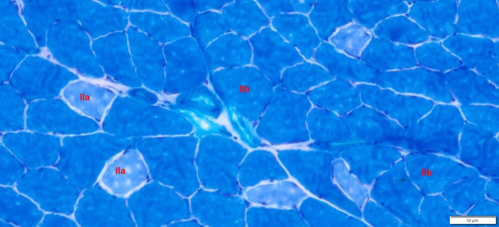
Expected Results
- Type I: dark blue
- Type IIa: pale blue
- Type IIb: medium blue
More Images
References
-
- Sawano S and Mizunoya W (2022). History and development of staining methods for skeletal muscle fiber types, Histol Histopathol. 37(6):493-503
- D’Angelis FH, et al (2014). Standardization of metachromatic staining method of myofibrillar ATPase activity of myosin to skeletal striated muscle of mules and donkeys. Pesq Vet Bras. 34(9):917-922.
- Ogilvie RW and Feeback DL (1990). A metachromatic dye-ATPase method for the simultaneous identification of skeletal muscle fiber types I, IIA, IIB and IIC. Stain Technology, 65(5), 231-241.
- Behan WMH, et al (2002).Validation of a simple, rapid, and economical technique for distinguishing type 1 and 2 fibres in fixed and frozen skeletal muscle. J Clin Pathol. 55(5): 375–380.
Note
This product is intended for research purposes only. This product is not intended to be used for therapeutic or diagnostic purposes in humans or animals.
Precautions
Handle with care. Avoid contact with eyes, skin and clothing. Do not ingest. Wear gloves.