Description
| SKU# | Organ | Pathology Diagnosis | Gender | Age | Grade | TMN | Thickness | IHC Data |
| HuPS-02004TA | Human Breast | Human breast Invasive ductal carcinoma and adjacent normal tissue | Female | 50 | 1 | 5μm | ER(100% +), PR(100%+), P53(+), CK5/6(-),P63(-), SMA(-),Ki67(25% +),Her2 (-),VEGF(+),E-cad(+) |
Slide Preparation
Human tissue was fixed in formalin immediately after excision and embedded in paraffin. The tissue sections were 5 µm in thickness and mounted each on positively charged glass slides.
Sections/slide
1 section on each slide
Slides/Package
10 slides (5 cancer and 5 normal tissue slides)
Applications
Histology stain, IHC/IF, FISH and ISH
Storage Condition
Store at 2-8ºC
H&E and IHC Image


Product Data Sheet and Material Safety Data Sheet (MSDS) (PDF)
Frequently Asked Questions
What is in this matched-pair package?
10 slides: 5 slides of human breast invasive ductal carcinoma and 5 slides of adjacent normal tissue, with 1 section on each slide.
Who was the donor?
A 50-year-old female. The tumor is grade 1 invasive ductal carcinoma.
What is the receptor and marker status of this tumor?
ER (100% +), PR (100% +), P53 (+), Her2 (-), CK5/6 (-), P63 (-), SMA (-), Ki67 (25% +), VEGF (+), and E-cad (+).
How were the sections prepared?
The human tissue was fixed in formalin immediately after excision and embedded in paraffin. Sections are 5 µm thick and mounted on positively charged glass slides.
What applications are these sections suitable for?
Histology staining, IHC/IF, FISH, and ISH.
What images are shown for this product, and how should the slides be stored?
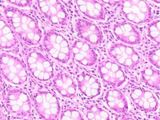
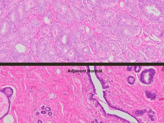
The page shows H&E images of the cancer and adjacent normal tissue at 2x, plus Ki67 IHC on both the cancer tissue and the normal breast tissue. Store at 2–8 ºC.