Description
Positive control slides are crucial in validating experimental techniques and ensuring the accuracy and reliability of staining procedures. In the context of Alzheimer’s disease research, brain sections from human patients with Alzheimer’s are commonly used as positive controls for assessing tau pathology and amyloid fibril deposition. The hallmark features of Alzheimer’s disease include the accumulation of hyperphosphorylated tau, forming neurofibrillary tangles, and the presence of amyloid plaques, both of which can be visualized through specific staining techniques.
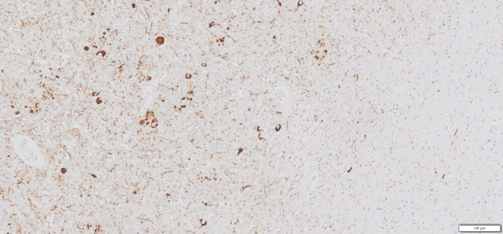
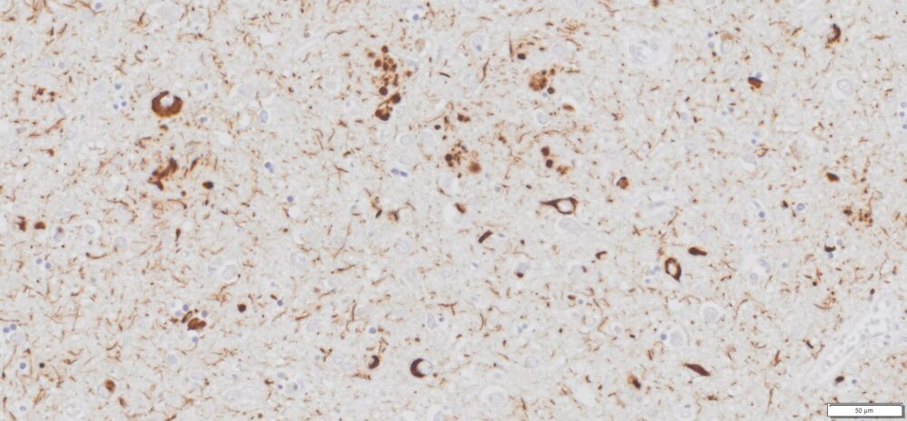
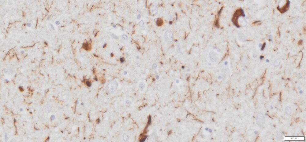
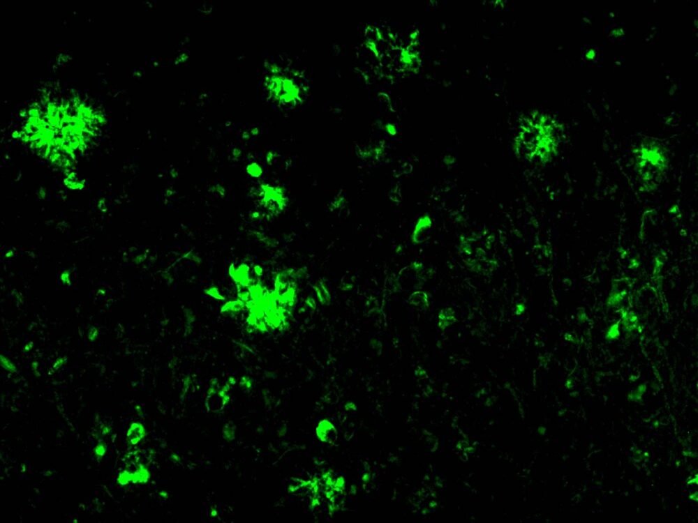
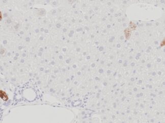
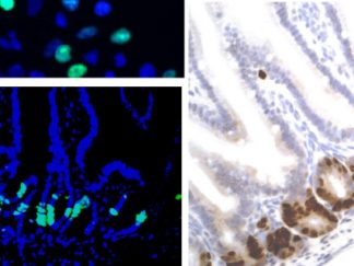
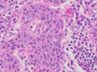
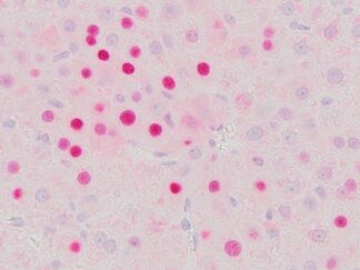
Tau immunohistochemistry (IHC/IF) enables the detection of phosphorylated tau, a key biomarker associated with neurodegeneration in Alzheimer’s disease. By utilizing antibodies that specifically recognize tau epitopes, this method reveals the presence, distribution, and severity of tau pathology in brain tissue. Thioflavin S staining, on the other hand, binds to beta-pleated sheet structures, such as those found in amyloid plaques, allowing for the visualization of amyloid deposits that are also critical to Alzheimer’s pathology.
In this study, positive control slides consisting of human Alzheimer’s disease brain sections have been prepared for both tau immunohistochemistry and Thioflavin S staining. These slides serve as a benchmark for assessing the performance of staining protocols and as a reference for comparison with other experimental samples. By ensuring the accuracy and consistency of these staining techniques, we aim to advance our understanding of Alzheimer’s disease pathology and improve diagnostic and therapeutic approaches.
Slide Preparation
Human brain tissue from an Alzheimer’s Disease autopsy was immediately fixed in formalin following excision and subsequently embedded in paraffin. The tissue sections, each 5 µm thick, were mounted onto positively charged glass slides.
Sections/slide
1 section on each slide
Slides/Package
5 slides per package
Applications
Tua IHC and Thioflavin S staining positive control
Storage Condition
Store at 2-8ºC
Images for Tau IHC and Thioflavin S Stain