Description
Alkaline phosphatase (ALP) is a membrane-associated enzyme widely expressed in tissues such as the liver, bone, kidney, and intestine. ALP participates in several important physiological processes, including bone mineralization, hepatic function, and intestinal nutrient absorption.
ALP activity is also widely used as a histochemical marker of cellular differentiation and tissue-specific enzyme activity. In stem cell research, elevated ALP activity is a characteristic feature of undifferentiated pluripotent stem cells, including embryonic stem cells (ESCs) and induced pluripotent stem cells (iPSCs). ALP activity generally decreases as cells undergo differentiation, making ALP histochemical staining useful for monitoring stem cell status and differentiation.
In tissue pathology, localized ALP activity can provide valuable information about enzyme distribution and cellular function. ALP staining may also help identify areas associated with phagocytic activity and inflammation in selected muscle biopsy specimens.
The VitroView™ Endogenous ALP Histochemical Staining Kit provides a convenient and reproducible method for detecting endogenous ALP enzymatic activity in cells and tissue sections. The staining reaction produces a localized colored reaction product at sites of ALP activity, allowing visualization by bright-field microscopy and providing qualitative or semi-quantitative assessment of enzyme activity.
Applications
- Stem Cell Research: Detection of ALP activity for screening and identifying undifferentiated pluripotent stem cells during stem cell culture, reprogramming, and differentiation studies.
- Histology and Cytochemistry: Visualization and localization of endogenous ALP activity in tissue structures, including selected muscle, vascular, hepatic, intestinal, and other ALP-expressing tissues.
- Pathology Research: Assessment of tissue-associated ALP activity and identification of localized areas of altered enzymatic activity in tissue specimens.
- Cell Biology: Evaluation of changes in ALP activity associated with cellular differentiation and functional changes.
Sample Compatibility
The kit is suitable for detecting endogenous ALP activity in 50–100 slides prepared from the following sample types:
- Cell smears
- Cultured cells
- Frozen tissue sections
Kit Contents
| VB-3048-1 | NewFuchsin Solution | 0.25 ml |
| VB-3048-2 | Sodium Nitrite Solution | 0.25 ml |
| VB-3048-3 | Naphthol AS-BI Solution | 0.25 ml |
| VB-3048-4 | ALP Buffer | 30 ml |
| VB-3048-5 | Mayer’s Hematoxylin Solution | 30 ml |
| VB-3048-6 | ALP Inhibitor Solution | 30 ml |
Storage
Store ALP Buffer, ALP Inhibitor Solution, and Mayer’s Hematoxylin Solution at room temperature. Store other reagents at -20°C away from light.
Procedure
- Sample Preparation:
- For Cell Smears (Blood / Bone Marrow): Prepare smears using fresh samples according to routine operations. Fix in ice-acetone for 2–3 minutes. Wash 3 times with distilled water. Proceed to Procedure Step 2.
- For Adherent Cells / Coverslips: Discard the culture medium completely. Wash 3–4 times with PBS. Fix in ice-acetone for 2–3 minutes. Wash 3 times with distilled water. Proceed to Procedure Step 2.
- For Frozen Sections: Warm the frozen sections to room temperature. Fix in ice-acetone for 2-3 minutes. Wash 3 times with distilled water. Proceed to Procedure Step 2.
- Staining
1) Prepare approximately 1.0 mL of New Fuchsin ALP staining working solution, sufficient for staining 2-5 slides, as follows:
- In a microcentrifuge tube, combine 10 μL of New Fuchsin Solution with 10 μL of Sodium Nitrite Solution. Incubate the mixture at room temperature for 1 min.
- Add 1.0 mL of ALP Buffer to the tube.
- Add 10 μL of Naphthol AS-BI Solution and mix thoroughly to prepare the working staining solution.
2) Use a hydrophobic barrier pen to draw a water-repellent circle around tissue sections or cells on the slide.
3) Gently drop the working solution to cover the cells or tissue section on the glass slides and incubate at room temperature for 30–60 min.
4) Drop Mayer’s Hematoxylin Solution onto the sections for 2–5 min; then wash the samples with running water.
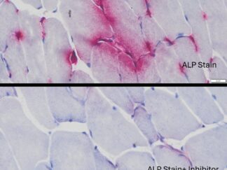
5) Specificity Control for ALP Activity (Option): Before step 3, apply ALP Inhibitor Solution to the slide and incubate for 30 minutes, wash with running water, then proceed to step 3.
6) Air-dry the slides and mount a coverslip onto the glass slide with VitroView™ Permanent Mounting Medium (SKU# VB-8003) or some other suitable organic mounting medium.
- Observation: Bright-field Microscopy can be used to examine specimens. When observing fluorescence, use a rhodamine excitation filter (500–570 nm).
Expected Results
Expected Results under Bright-field Microscopy
- ALP Positive —————wine-red
- Nuclei———————– light blue
Expected Results under Fluorescence Microscopy
- ALP Positive ——— red
Positive Controls
- Mouse or human Muscle or kidney frozen section
Refeence
- Zhou L, et al (2026). Simultaneous coupling azo dye methods for diagnosing capillarization: Standardizing endogenous alkaline phosphatase histochemistry on fresh frozen striated muscle biopsy specimens. Journal of Neuromuscular Pathology, 18(3): 214–223
More ALP Staining Images


Precautions:
- Handle reagents with care.
- Avoid contact with eyes, skin, or clothing.
- Do not ingest.
- Always wear gloves when handling chemicals.