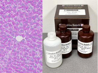
Description
The VitroSure™ DNA FFPE Tissue Isolation Kit delivers fast, reliable, and high-yield DNA extraction from formalin-fixed, paraffin-embedded (FFPE) tissue samples. Engineered for both genomic and mitochondrial DNA isolation, this kit features proprietary VitroSure™ DNA Elute Columns and advanced silica membrane technology for consistent purification results. With flexible elution volumes (20–100 µl) and optimized performance for small tissue samples, this cost-effective, ready-to-use kit is ideal for sensitive downstream applications, including PCR, qPCR, NGS, and genotyping.
Technical Specifications
| Equipment needed | Microcentrifuge, heat block/bath (37°C, 56°C and 90°C) |
| DNA Type Isolated | Total DNA |
| Size Range | > 50 bp |
| Yield | Up to 25 µg total DNA can be eluted into ≥ 50 µl |
| Purity | Typical A260/A280 ≥ 1.8 |
| Eluted DNA Storage | at ≤ -20°C |
| Sample Source | Tissue from a paraffin block or tissue sections |
| Processing Capacity | FFPE Tissue: ≤25 mg or 2-8 sections at a thickness of 7-10 µm with
a surface area of 15-20 mm2 |
| Applicable For | PCR and Next Generation Sequencing (NGS), genotyping,
Restriction enzyme digestion, SNP, etc. |
Kit Contents
|
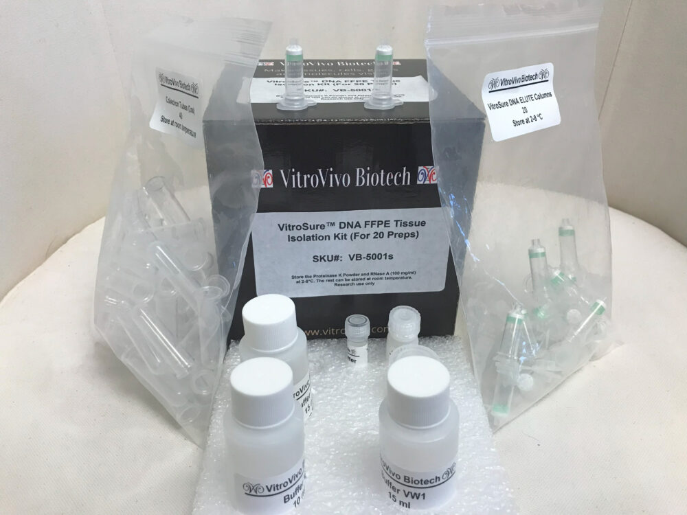 |
Storage
Store the Proteinase K Powder and RNase A (100 mg/ml) at 2-8°C. After reconstitution of Proteinase K, store the solution at -20°C. The rest can be stored at room temperature.
Procedures
- Sample preparation
1) Sample preparation from FFPE block using the Deparaffinization Solution (VB-5009, sold separately):
- Use a scalpel to trim excess paraffin off the sample block.
- Cut up to 2-6 sections with a microtome, each 5–10 µm thick. The section number for each sample depends on the tissue size. Discard the first 2–3 sections if the sample surface has been exposed to air.
- Place the sections immediately in a 1.5 or 2 ml microcentrifuge tube
- Add Deparaffinization Solution: for 2-6 sections or one 20 µm section, add 320 µl Deparaffinization Solution; for more sample material, add 640 µl Deparaffinization Solution.
- Vortex vigorously for 10 s, and centrifuge briefly to bring the sample to the bottom of the tube.
- Incubate at 56°C for 3 min, then allow to cool at room temperature (15–25°C), and centrifuge at full speed for 2 min.
- Carefully remove the supernatant by pipetting without disturbing the pellet. Carefully remove any residual Deparaffinization Solution using a fine pipette tip.
- Keep the lid open and incubate for 10 min at 37°C to dry the pellet. Proceed to step 3
2) Sample preparation from FFPE block using Xylene:
- Use a scalpel to trim excess paraffin off the sample block.
- Cut up to 2-6 sections with a microtome, each 5–10 µm thick. The section number for each sample depends on the tissue size. Discard the first 2–3 sections if the sample surface has been exposed to air.
- Place the sections immediately in a 1.5 or 2 ml microcentrifuge tube and add 1 ml of xylene to the sample. Close the lid and vortex vigorously for 10 seconds.
- Centrifuge at maximum speed for 2 minutes at room temperature.
- Carefully remove the supernatant without disturbing the pellets.
- Add 1 ml of ethanol (96–100%) to the pellet and mix by vortexing to extract residual xylene from the sample.
- Centrifuge at maximum speed for 2 minutes at room temperature.
- Carefully remove the supernatant without disturbing the pellet. Remove any remaining ethanol with a fine pipette tip.
- Open the tube and incubate at room temperature or up to 37°C for 10minutes or until all residual ethanol has evaporated. Proceed to step 3
3) Sample preparation from FFPE sections on slides:
- Submerge the slides in xylene I for 3 minutes, followed by xylene II for an additional 3minutes.
- Remove xylene by rinsing with 100% ethanol (1 minute each, repeated twice).
- Air dry the slides for 3-5minutes.
- Gently detach the tissue sections from the slides using a small blade, then transfer the tissue pellets into a 1.5 ml microcentrifuge tube.
- Proteinase K solution preparation: Combine 260 µl of Proteinase K Buffer with 5 mg of Proteinase K Powder. Vortex the mixture to ensure complete dissolution. Store the solution at -20°C.
- Resuspend the pellets in 180 µl Buffer VTL. Add 20 µl of proteinase K solution and mix by vortexing.
- Incubate at 56°C for 1 hour or until the sample is completely lysed.
- Incubate at 90°C for 1 hour without agitation.
- Cool to room temperature and briefly centrifuge the tube. For RNA-free genomic DNA, add 2 µl of RNase A (100 mg/ml) and incubate at room temperature for 2 minutes.
- Add 200 µl of Buffer VL and 200 µl ethanol to the sample. Mix thoroughly by vortexing.
- CarefullytransfertheentirelysatetoaDNAElutecolumnandcentrifugeat8000×g (or 10000 rpm) for 1 minute. Discard the flow-through.
- Add 500 µl of Buffer VDW1 and centrifuge at 8000×g (or 10000 rpm) for 1 minute. Discard the flow-through.
- Add500µlofBufferVDW2andcentrifugeat8000×g (or10000 rpm) for1 minute. Discard the flow-through and collection tube.
- Place the DNA Elute column in a clean 2 ml collection tube. Centrifuge at maximum speed for 3 minutes with the lid open to completely dry the membrane.
- 5ml microcentrifugetubeandapply20–100µl of Buffer VTE to the center of the membrane. Ensure that Buffer VTE is at room temperature.
- Incubate at room temperature for 5 minutes and centrifuge at maximum speed (20,000×g or 14,000 rpm) for 1 minute to elute the DNA.
User Manual and Material Safety Data Sheet (MSDS) (PDF )
Note
This product is intended for research purposes only. This product is not intended to be used for therapeutic or diagnostic purposes in humans or animals.
Precautions
Handle with care. Avoid contact with eyes, skin and clothing. Do not ingest. Wear gloves.