Description
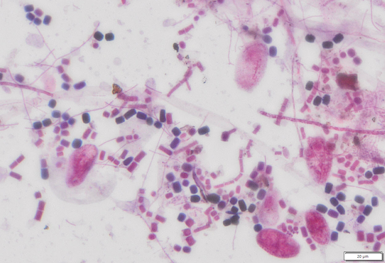
The Gram stain is fundamental to the phenotypic characterization of bacteria. The staining procedure differentiates organisms of the domain Bacteria according to cell wall structure. Gram-positive cells have a thick peptidoglycan layer and stain blue to purple. Gram-negative cells have a thin peptidoglycan layer and stain red to pink. This kit is designed for the gram staining of slide smear sample.
Kit Contents
| VB-2000-1 | Gram’s Crystal Violet Solution | 250 ml |
| VB-2000-2 | Gram’s Iodine Solution | 250 ml |
| VB-2000-3 | Gram’s Decolorizer Solution | 250 ml |
| VB-2000-4 | Gram’s Safranin Solution | 250 ml |
Storage
Room temperature.
Procedure
Prepare a Slide Smear:
- Use a glass etching tool to mark one or more dime-sized circles on the surface of a slide.
- Transfer a drop of the suspended culture to be examined on a slide with an inoculation loop. If the culture is to be taken from a Petri dish or a slant culture tube, first add a drop or a few loopful of water on the slide and aseptically transfer a bit of the colony. It should only be a very small amount of culture. A visual detection of the culture on an inoculation loop already indicates that too much is taken.
- Spread the culture with an inoculation loop to an even thin film over a circle of 1.5 cm in diameter.
- It is possible to put 3 to 4 small smears on a slide, if more than one culture is to be examined.
- Hold the slide with a clothes-pin. Allow to air dry and fix it over a gentle flame, while moving the slide in a circular fashion to avoid localized overheating. The applied heat helps the cell adhesion on the glass slide to make possible the subsequent rinsing of the smear with water without a
Gram Staining
- Flood fixed smear of cells with Gram’s Crystal Violet solution. Please note that the quality of the smear (too heavy or too light cell concentration) will affect the Gram Stain results.
- Wash slide in a gentle and indirect stream of tap water for 2 seconds.
- Flood with Gram’s iodine Solution. Allow it to remain for Gram’s Crystal Violet Solution.
- Pour off the iodine solution and gently wash with tape water. Shake off the excess water from the surface.
- Decolorize with Gram’s Decolorizer Solution until the blue dye no longer flows from the smear. Further delay will cause excess decolorization in the gram-positive cells, and the purpose of staining will be defeated.
- Gently wash the smear with tape water.
- Flood slide with counterstain, Gram’s safranin Solution. Wait 30 seconds to 1 minute.
- Wash off the red safranin solution with water. Blot with bibulous paper to remove the excess water. Alternatively, the slide may be shaken to remove most of the water and air-dried.
- Observe the results of the staining procedure under oil immersion using a bright field microscope.
Result
- Gram-positive organisms——————bluish purple
- Gram-negative organisms—————–pinkish red
Note
This product is intended for research purposes only. This product is not intended to be used for therapeutic or diagnostic purposes in humans or animals.
Precautions
Handle with care. Avoid contact with eyes, skin and clothing. Do not ingest. Wear gloves.

