Description
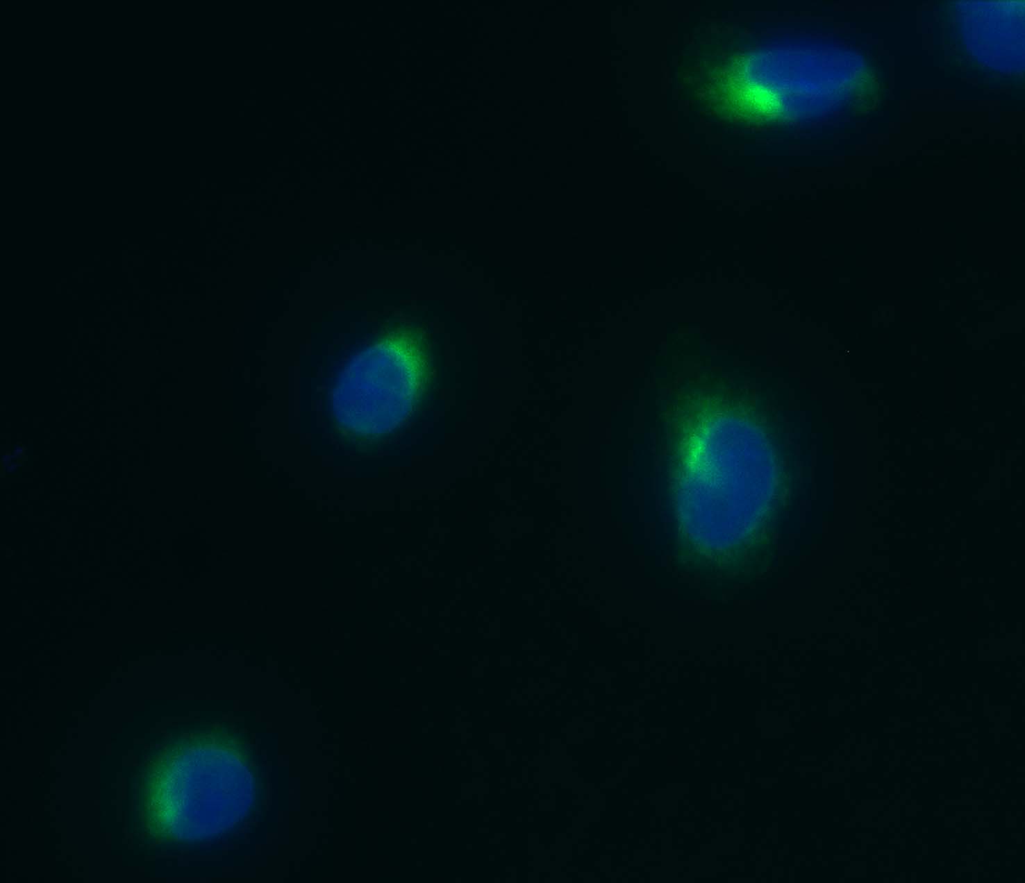
The endoplasmic reticulum (ER) is an organelle found in most eukaryotic cells, that forms an interconnected network of flattened, membrane-enclosed sacs or tubes. The ER has a central role in lipid and protein synthesis, protein chaperoning and folding, and calcium homeostasis. DiOC6(3) is a cell-permeant, green-fluorescent (EX:482nm/EM:504nm), lipophilic dye that is selective for the mitochondria of live cells, when used at low concentrations. At higher concentrations, the dye may be used to stain other internal membranes, such as the endoplasmic reticulum. VitroVivo Biotech offers a ready -to-use (RTU) cell-permeant probe DiOC6(3) that selectively stain the ER in live cells based on the probe concentration.
Content
VB-1004 RTU Endoplasmic Reticulum Staining Solution——–30 ml
Storage Condition
Store at 2-8 °C and protect from light.
Application
Cell endoplasmic reticulum (ER) staining
General Protocol:
Adherent cells for fluorescence microscopy
- Grow cultured cells on sterile glass cover slips or slides overnight at 37 ºC.
- Follow appropriate protocol to fix cultured cells.
- Completely wash the cells with PBS as needed.
- Add adequate RTU Endoplasmic Reticulum Staining Solution to cover the whole sample.
- Incubate under dark at room temperature for 15-30 minutes.
- Rinse the sample several times with PBS and remove excess dye.
- Add antifade aqueous mounting medium and mount.
- Use appropriate filters and detect under fluorescence microscope according to standard protocol.
Suspension cells for fluorescence microscopy
- The cells are harvested into a 15 mL polypropylene centrifuge tube and spin down for 8 min at 600 RPM,
- The supernatant is discarded and the cells are resuspended in 0.5 ml of culture medium
- 1-2 drops of the cell suspension were placed on a slide in the central area and moved around to form a thin and even film with a glass spreader.
- (Option) You also can use cytocentrifuge to prepare cell slides.
- Air dry and follow appropriate protocol to fix cultured cells.
- Drop adequate RTU Endoplasmic Reticulum Staining Solution to cover the whole sample on slide.
- Incubate under dark at room temperature for 15–30 minutes.
- Cover with coverslip and view under fluorescence microscope according to standard protocol.
Note: This product is intended for research purposes only. This product is not intended to be used for therapeutic or diagnostic purposes in humans or animals.
References
- Soldani C, et al. Apoptosis in tumour cells photosensitized with Rose Bengal acetate is induced by multiple organelle photodamage.Histochem Cell Biol (2007) 128:485-495
- Foissner I. Fluorescent phosphocholine–a specific marker for the endoplasmic reticulum and for lipid droplets in Chara internodal cells.Protoplasma (2009) 238:47-58
- Leys N, et al. The response of Cupriavidus metallidurans CH34 to spaceflight in the international space station. Antonie Van Leeuwenhoek (2009) 96:227-245