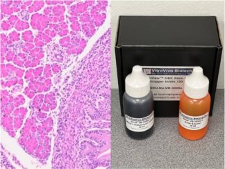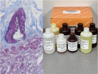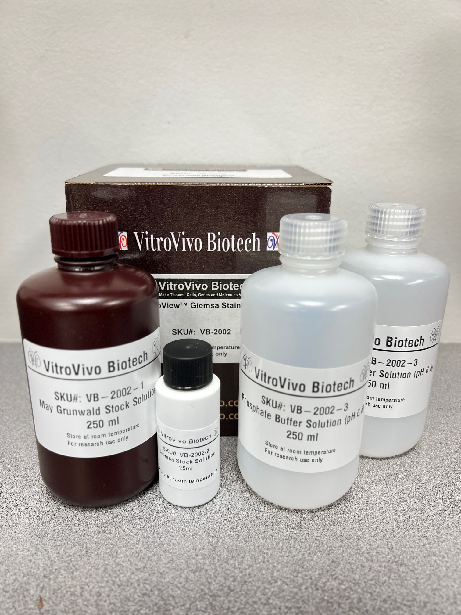
Description
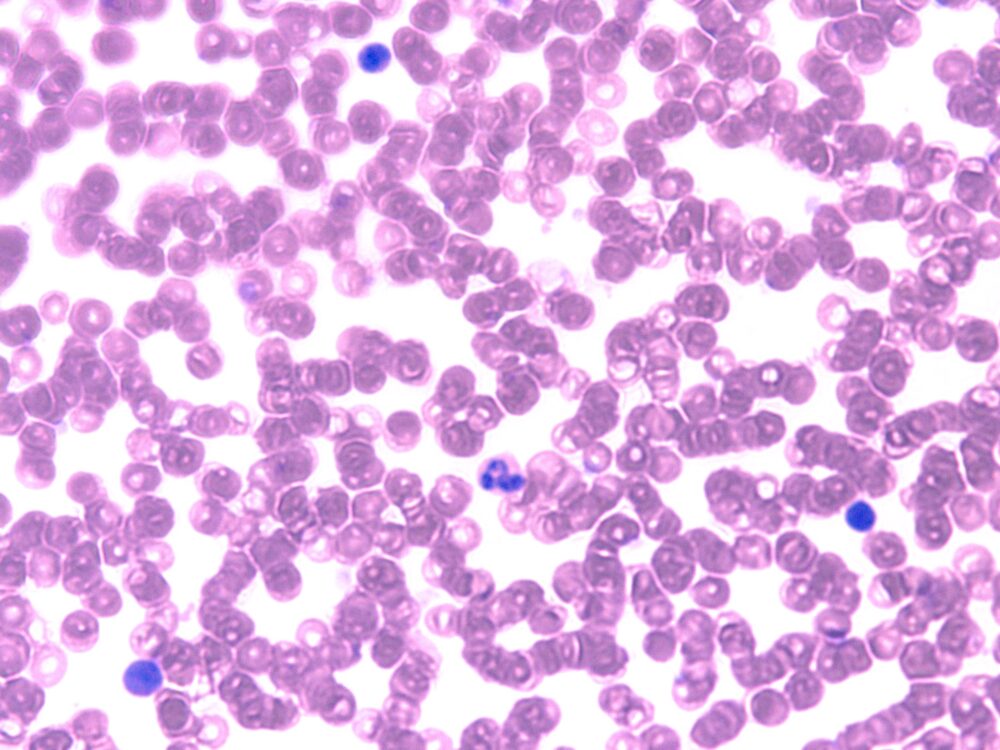
VitroView™ Giemsa Stain Kit (May Grunwald) is intended for use in the visualization of cells present in hematopoietic tissues and certain microorganisms. This kit may be used on blood smear, formalin-fixed, paraffin-embedded or frozen sections.
Kit Contents
| VB-2002-1 | May Grunwald Stock Solution | 250 ml |
| VB-2002-2 | Giemsa Stock Solution | 25 ml |
| VB-2002-3 | Phosphate Buffer Solution (pH6.8) | 250 ml×2 |
Storage
Room temperature.
Procedure
Reagent Preparation:
- Prepare Working May-Grunwald Solution by mixing 25 ml of May-Grunwald Solution with 25 ml of PBS Solution, pH 6.8
- Prepare Working Giemsa Solution by mixing 2.5 ml of Giemsa Stock Solution with 50 ml of PBS, pH 6.8
Standard Procedure:
- Deparaffinize sections if necessary and hydrate to distilled water.
- Place slide in staining tray and flood with Working May-Grunwald Solution for 5-7 minutes. Agitate slide occasionally to insure proper staining.
- Carefully flood slide with Phosphate Buffer Solution, pH 6.8 until stain no longer runs off.
- Flood slide with Working Giemsa Solution for 10-15 minutes. Note: Agitate slide occasionally to insure proper staining.
- Carefully flood slide with Phosphate Buffer Solution, pH 6.8 until stain no longer runs off.
- Allow slide to remain in Phosphate Buffer Solution, pH 6.8 for an additional 3 minutes.
- Dip slide quickly in distilled water to remove buffer.
- Dehydrate in 5 dips each of 95% and 100% ethyl alcohol. Clear in three changes of xylene, 3 minutes each.
- Mount coverslip onto glass slide with Permount or some other suitable organic mounting medium
Procedure for Mast Cells:
- Deparaffinize sections if necessary and hydrate to distilled water.
- Place slide in staining tray and flood with Working May-Grunwald Solution for 5-7 minutes. Note: Agitate slide occasionally to insure proper staining.
- Carefully flood slide with Phosphate Buffer Solution, pH 6.8 until stain no longer runs off.
- Flood slide with Working Giemsa Solution for 10-15 minutes. Note: Agitate slide occasionally to insure proper staining.
- Carefully flood slide with Phosphate Buffer Solution, pH 6.8 until stain no longer runs off.
- Differentiate by dipping slide in Acetic Acid Solution (0.25%) until background is desired intensity.
- Dip slide for 10 seconds in Phosphate Buffer Solution, pH 6.8 while agitating gently.
- Dip slide quickly in distilled water to remove buffer and air dry at room temperature.
- Dehydrate in 5 dips each of 95% and 100% ethyl alcohol. Clear in three changes of xylene, 3 minutes each.
- Mount coverslip onto glass slide with Permount or some other suitable organic mounting medium
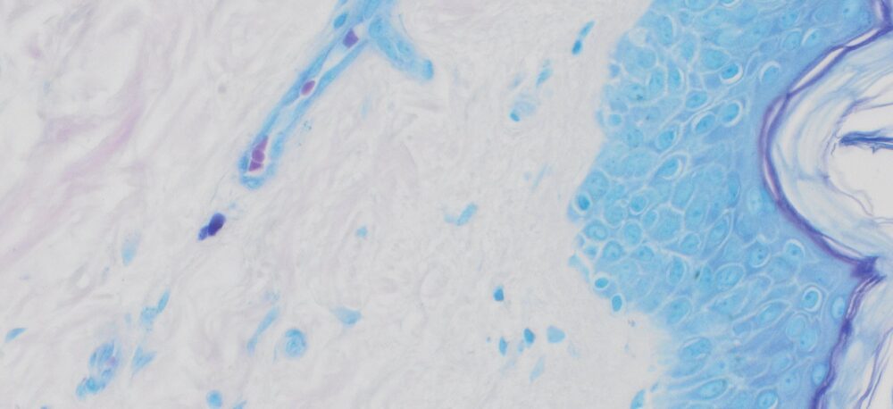
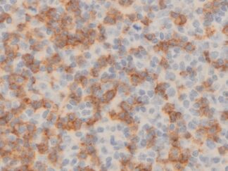
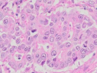
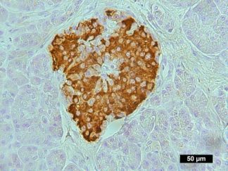
Expected Results
- Nuclei—————————————-Blue/Violet
- Cytoplasm———————————–Light Blue
- Collagen————————————-Pale Pink
- Muscle Fibers——————————-Pale Pink
- Erythrocytes———————————Gray, Yellow or Pink
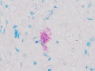
- Rickettsia————————————Reddish-Purple
- Helicobacter pylori————————Blue
- Mast Cells———————————–Dark Blue with Red Granules
Control Tissue
Blood film; Bone Marrow; Spleen; or any well fixed tissue.
Note
This product is intended for research purposes only. This product is not intended to be used for therapeutic or diagnostic purposes in humans or animals.
Precautions
Handle with care. Avoid contact with eyes, skin and clothing. Do not ingest. Wear gloves.
User Manual and Material Safety Data Sheet (MSDS) (PDF)
More Images