Description
Tartrate-Resistant Acid Phosphatase (TRAP) is an acid phosphatase enzyme that remains active in the presence of tartrate. The enzyme hydrolyzes a chromogenic substrate, producing a colored reaction product at sites of TRAP activity.
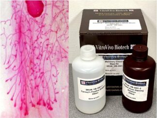
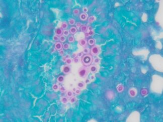
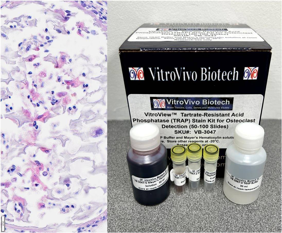
TRAP-positive cells typically appear as red, dark red, or purple red, depending on the kit formulation and counterstain used. Osteoclasts are generally identified as multinucleated TRAP-positive cells.
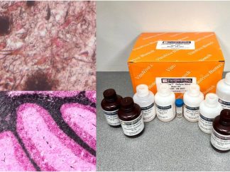
This VitroView™ Tartrate-Resistant Acid Phosphatase (TRAP) Stain Kit is designed for histochemical identification of osteoclasts and other TRAP-positive cells in tissue sections and cell preparations.
Key Advantages
- Dual-Modality Imaging: Enables both bright-field and fluorescence microscopy evaluation.
- Organic Mounting Compatible: Formulated with NewFuchsin to withstand alcohol dehydration and xylene clearing.
- High Yield Capacity: Single kit processes 50 to 100 slides efficiently.
- Versatile Sample Compatibility: Validated for cell smears, adherent cells, frozen sections, and paraffin sections.
- Streamlined Working Protocol: Micro-volume master mix preparation takes less than two minutes. Clear Cellular Contrast: Delivers distinct wine-red cytoplasm against light blue nuclei.
Application
- Identification of osteoclasts in bone tissue
- Assessment of osteoclast differentiation in cell cultures
- Research applications involving bone remodeling and resorption
- Histological and cytochemical investigations
Kit Contents
| VB-3047-1 | NewFuchsin Solution | 0.25 ml |
| VB-3047-2 | Sodium Nitrite Solution | 0.25 ml |
| VB-3047-3 | Naphthol AS-BI Solution | 0.25 ml |
| VB-3047-4 | TRAP Buffer | 30 ml |
| VB-3047-5 | Mayer’s Hematoxylin Solution | 30 ml |
Storage
Store TRAP Buffer and Mayer’s Hematoxylin solution at room temperature away from light. Store other reagents at -20°C away from light. This kit is stable for at least 3 months.
Procedure
- Sample Preparation:
- For Cell Smears (Blood / Bone Marrow): Prepare smears using fresh samples according to routine operations. Fix in 10% Neutral Buffered Formalin (NBF) for 15–30 minutes. Wash 3 times with distilled water. Proceed to Procedure Step 2.
- For Adherent Cells / Coverslips: Discard the culture medium completely. Wash 3–4 times with PBS. Fix in 10% Neutral Buffered Formalin (NBF) for 15–30 minutes. Wash 3 times with distilled water. Proceed to Procedure Step 2.
- For Frozen Sections: Warm the frozen sections to room temperature. Fix in 10% Neutral Buffered Formalin (NBF) for 15–30 minutes. Wash 3 times with distilled water. Proceed to Procedure Step 2.
- For Paraffin Sections: Deparaffinize sections in xylene (2 × 6 min), followed by rehydration in 100% ethanol (2 min), 95% ethanol (2 × 2 min), and 70% ethanol (2 min). Rinse in distilled water for 5 min and proceed to Procedure Step 2.
- Staining
- Prepare approximately 1.0 mL of New Fuchsin TRAP staining working solution, sufficient for staining 2-5 slides, as follows:
- In a microcentrifuge tube, combine 10 μL of NewFuchsin Solution with 10 μL of Sodium Nitrite Solution. Incubate the mixture at room temperature for 1 min.
- Add 1.0 mL of TRAP Buffer to the tube.
- Add 10 μL of Naphthol AS-BI Solution and mix thoroughly to prepare the working staining solution.
- Use hydrophobic barrier pen to draw a water-repellent circle around tissue sections or cells on the slide.
- Gently drop the working solution to cover the cells or tissue section on the glass slides and incubate at 37 °C in moisture chamber for 40–60 min.
- Drop Mayer’s Hematoxylin Solution onto the bone sections for 2–5 min; then wash the samples with running water for 15 min.
- Dehydration and mounting
- Dehydrate with 2 changes of 95% Ethanol and 2 changes of 100% Ethanol (2 minutes per change).
- Clear with 3 changes of xylene (5 minutes per change)
- Mount coverslip onto glass slide with Permount or some other suitable organic mounting medium.
- Observation: Bright-field Microscopy can be used to examine specimens. When observing fluorescence, use a rhodamine excitation filter (500–570 nm).
Expected Results
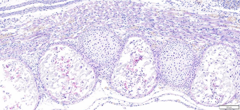
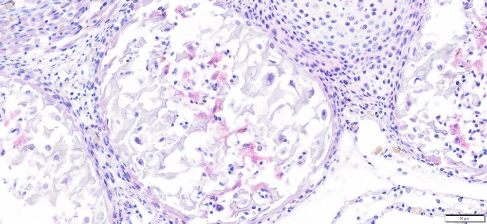
Expected Results under Bright-field Microscopy
- Osteoclast cytoplasm —-wine-red
- Nuclei———————– light blue
Expected Results under Fluorescence Microscopy
- Osteoclast cytoplasm ——— red
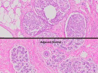
Positive Controls
- Mouse fetus whole sections (Spinal bone)
- Metaphyses or growth plates from juvenile mice or rats (3 to 6 weeks old) are the most common laboratory controls.
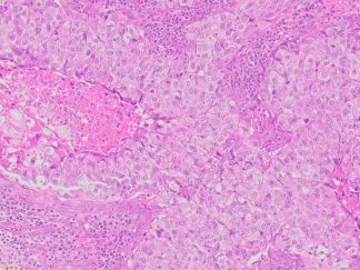
- Human giant cell tumor tissue sections
References
- Nakamura A, et al (2025). Osteoclast visualization: Tartrate-resistant acid phosphatase activity staining using NewFuchsin compatible with non-aqueous mounting and tissue clearing. Methods X, 14: 103136
- Luo G, et al (2025). Precision-targeting and dual silencing osteoclastogenesis and inflammatory pathways for the treatment of radiation-induced bone deterioration. Biomaterials Advances, 117:
More Tartrate-Resistant Acid Phosphatase (TRAP) Staining Images
Precautions:
- Handle reagents with care.
- Avoid contact with eyes, skin, or clothing.
- Do not ingest.
- Always wear gloves when handling chemicals.