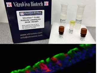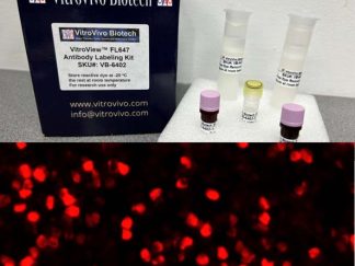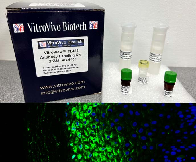
Description
The VitroView™ FL488 Antibody Labeling Kit provides a highly efficient workflow for conjugating IgG antibodies with FL488 dye. The kit utilizes amine-reactive NHS esters to form a stable, covalent bond with the antibody’s primary amines. Ready-to-use dye-removal columns are included to separate the labeled antibody from excess unreacted fluorophore, consistently achieving final yields above 90%.
Key Advantages
- High Signal-to-Noise Ratio: Deliver superior results with minimal background fluorescence and reduced non-specific binding.
- Rapid & User-Friendly: Optimized protocol ensures minimal hands-on time and highly reproducible results.
- Complete System: Contains essential primary reagents required for successful fluorophore conjugation.
- Streamlined Workflow: Simple, direct steps minimize potential handling errors and save valuable bench time.
Specifications
| Product | Molecular Weight | Ex/Em | Molar extinction coefficient (cm-1M-1) | CF280 |
| FL488 | 631.6 | 499/520 | 73,000 | 0.11 |
Kit Contents
| SKU | Contents | Amount |
| VB-6400-1 | FL488 Reactive Dye | 2 vials |
| VB-6400-2 | Reactive Buffer (10×) | 50 µL |
| VB-6400-3 | Free dye Removal Column | 2 Unit |
Storage
Store reactive dye at -20 °C immediately upon receipt.
Reagents and Materials Required but Not Provided
- Microcentrifuge capable of 5,000 × g
- 10 mM phosphate-buffered saline (PBS), pH 7.4
- Amicon Ultra-4 Centrifugal Filter (Millipore)
Protocol
- Prepare your antibody before using a labeling kit.
1) Pre-Labeling Requirements
- Purity: Antibody must be >90% pure.
- Concentration: Ideal range is 0.5–2.0 mg/mL.
- Formulation: Must be completely free of amine-containing additives and carrier proteins.
2) Common Interfering Substances
| Substance | Maximum Allowed | Why It Interferes | Solution |
| Tris / Glycine | 0 mM | Competes for binding sites | Dialysis / Buffer exchange |
| Sodium Azide | <0.02% (Ideally 0%) | Inhibits certain reactions | Dialysis / Buffer exchange |
| BSA / Gelatin | 0% | Labels the carrier protein instead | Protein A/G or other affinity purification |
| Glycerol | <10% | Reduces labeling efficiency | Dialysis / Buffer exchange |
| Ammonium ions | 0 mM | Competes for reactive groups | Dialysis / Buffer exchange |
3) Dialysis or buffer exchange your antibody if your antibody contains Tris / Glycine, Sodium Azide, Glycerol or Ammonium ions; we suggest using a centrifugal spin filter (e.g., Amicon Ultra).
- Choose an appropriate Molecular Weight Cut-Off (MWCO): For IgG (150 kDa): Use a 10 kDa or 30 kDa MWCO filter.
- Add 500 µL of compatible buffer (e.g., 1X PBS, pH 7.4).
- Spin at 14,000 × g for 5 minutes.
- Discard the flow-through.
- Load the antibody sample into the filter.
- Top up with PBS to the maximum volume line.
- Spin according to manufacturer speed recommendations.
- Discard flow-through; repeat this wash step 3 times.
- Invert the spin column into a clean collection tube.
- Spin at 1,000 × g for 2 minutes to collect the purified antibody.
- Concentration Verification: UV Absorbance: Measure at A280 using a spectrophotometer. IgG Extinction Coefficient: Use 1.4 for a 1 mg/mL solution. Calculation: Concentration (mg/mL) = A280 / 1.4.
4) Purify your antibody if your antibody contains BSA / Gelatin
To separate BSA or gelatin from your antibody, you must use a method based on chemical properties or affinity rather than simple size-exclusion filtration:
- Negative Selection Resins: Commercial kits like the Thermo Scientific Melon Gel IgG Purification System bind to BSA and gelatin while allowing the pure antibody to flow straight through.
- Dedicated BSA Removal Kits: Specialized kits such as the Abcam BSA Removal Kit (ab173231) chemically precipitate or capture BSA out of the solution specifically for downstream conjugation.
- Protein A, G, or L Affinity Chromatography: This method binds your antibody tightly to a resin while you wash the BSA and gelatin away, allowing you to elute the pure antibody afterward.
5) Adjust the antibody concentration to 1 mg/mL. If the antibody concentration is ≥1 mg/mL, dilute it to 1 mg/mL with PBS; if it is <1 mg/mL, concentrate the sample by centrifugation until it reaches 1 mg/mL.
6) For Lyophilized Antibody: Reconstitute the lyophilized antibody powder in this diluted buffer to achieve a final concentration of 1 mg/mL.
2. Antibody Labeling Reaction
- Mix the Reactive buffer (10×) with the antibody solution at a ratio of 1:10
- Transfer 110 µL of the prepared 1 mg/mL antibody solution directly into one vial of FL488 reactive dye.
- Cap the vial securely and gently invert it several times to completely dissolve and mix the dye with the antibody.
- Incubate the reaction mixture for 1 hour at room temperature in the dark.
3. Free Dye Removal & Purification
- Remove the column cap and pour off the storage buffer. Cut the sealed bottom end of the column at the marked notch.
- Equilibrate the column: Fill the column with PBS (pH 7.4), allowing the buffer to gravity-flow completely through the packed bed. Repeat this step 4 times, discarding all flow-through.
- Dilute the reaction mixture: Add PBS to your 100 µL labeled antibody solution to bring the final volume up to 2.5 mL.
- Load the 2.5 mL sample carefully onto the top of the equilibrated column bed.
- Allow the sample to completely enter the packed bed by gravity flow. Discard the resulting flow-through.
- Elute the purified antibody: Place a clean collection tube underneath the column and add 3.5 mL of PBS buffer to elute the conjugate.
4. Optional: Antibody Concentration
If required, concentrate the eluted antibody to the desired working concentration using a 10-kDa or 30-kDa molecular-weight cutoff Amicon® centrifugal filter unit (not provided).
5. Optional: efficiency test of labeling reaction
- Calculate antibody concentration C (mg/ml) and dye/antibody degree of labeling (DOL) by measuring OD280 and OD496 absorbance:
C = (MWAb × [OD280-CF280 × OD496]) / εAb
DOL = (OD496×εAb) / (εdye × [OD280 – CF280 ×OD496])
MWAb: Molecular weight of antibody (150 kDa)
OD280: Absorbance at 280nm of the dye-antibody conjugate
OD496: Absorbance at 496nm of dye
CF280: Correction factor at 280nm accounting for absorbance of dye at 280nm (0.11)
εAb: Molar extinction coefficient at 280nm of antibody (210,000 cm-1M-1)
εdye: Molar extinction coefficient at 496nm of the dye (73,000 cm-1M-1)
- Make final concentration at 1mg/ml in PBS buffer with 0.1% BSA, 0.02% Na3N (option).
Note
This product is intended strictly for laboratory research purposes. It is not approved for therapeutic, clinical, or diagnostic procedures in humans or animals.
Precautions
Handle with care. Avoid contact with eyes, skin, and clothing. Do not ingest. Wear gloves.
User Manual and Material Safety Data Sheet (MSDS) (PDF)
VB-6400 MSDS