Histochemical Stains
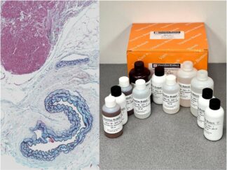
VitroVivo Biotech Provides Histochemical Staining kits for Tissue and Cell Visualization
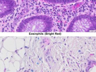
Histochemical stains are essential for visualizing tissue and cellular structures under a microscope. Since living or fixed cells have minimal natural color and only slight differences in refractive indices, staining enhances contrast, allowing scientists and pathologists to study tissue morphology and detect specific cell types, structures, or microorganisms.
Types of Histochemical Stains
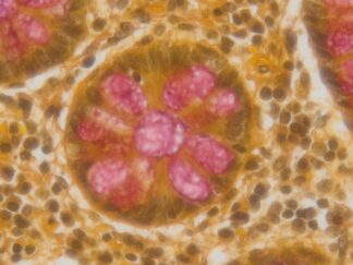
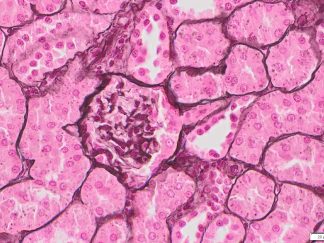
- Routine Stains: Hematoxylin and eosin (H&E stain) is the most widely used technique for general tissue structure visualization.
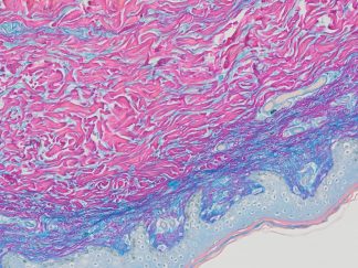
- Special Stains: These methods highlight specific tissue components, elements, or microorganisms that H&E staining cannot detect.
Explore Our Histochemical Stain Kits
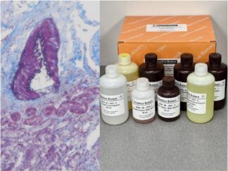
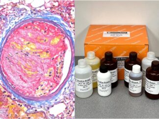
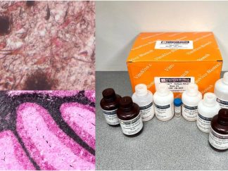
At VitroVivo Biotech, we offer over 36 high-quality histochemical staining kits designed for both routine and special staining applications. Browse our selection below to find the right stain kit for your research or diagnostic needs.
Guidance on Choice of VitroView™ Histochemical Stain Kits
| Product Name | SKU# | Visualization For |
| Hematoxylin and Eosin Stain Kit | VB-3000 | General morphology of tissues and cells |
| VB-3001 | Structure of the whole mouse mammary glands | |
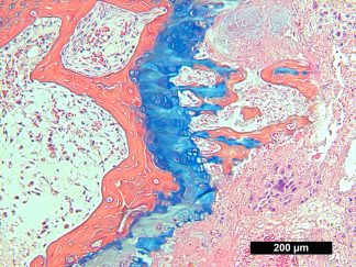
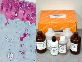
| Alcian Blue Hematoxylin-Orange G Stain Kit | VB-3002 | Cartilage, mature bone and immature bones |
| VB-3003 | Tissue mucosubstances | |
| PAS Stain Kit | VB-3004 | Glycogen, mucin and fungi |
| VB-3005 | Both acidic and neutral mucins as well as mixtures of acidic and neutral mucins | |
| VB-3006 | Basic neuronal structure in brain or spinal cord tissues | |
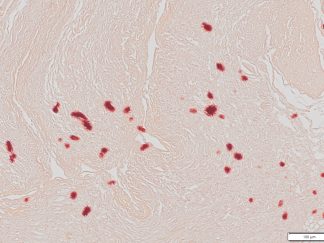
| Oil Red O Stain Kit | VB-3007 | Lipid and fat staining on frozen sections |
| Alizarin Red Stain Kit | VB-3008 | Calcium on tissue sections |
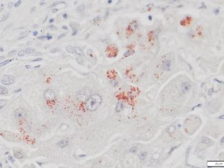
| Prussian Blue Stain Kit | VB-3009 | Ferric iron in tissue sections |
| Nissl Stain Kit | VB-3010 | Nissl body in the cytoplasm of neurons |
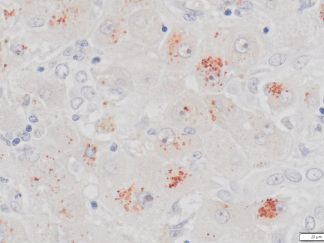
| VB-3011 | Amyloid deposits | |
| VB-3012 | Lipid and fat staining on frozen sections | |
| Toluidine Blue Stain Kit | VB-3013 | Mast cells |
| VB-3014 | Connective fibers | |
| VB-3015 | Nerve fibers, neurites and neurofibrillary tangles | |
| VB-3016 | Collagen and muscle | |
| VB-3017 | Collagen fibers | |
| VB-3018 | Reticular fibers | |
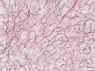
| VB-3019 | Elastic fibers | |
| VB-3020 | Argentaffin granules and melanin | |
| Rhodanine Copper Stain Kit | VB-3021 | Copper deposits |
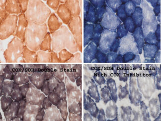
| COX/SDH Double Histochemistrical Stain Kit | VB-3022 | Detection of mitochondrial function |
| Movat Pentachrome Stain Kit | VB-3023 | Collagen, muscle tissue, reticular fibers, mucins and fibrin |
| Southgate’s Mucicarmine Stain Kit | VB-3024 | Mucin |
| Jones Stain Kit | VB-3026 | Basement membrane |
| Colloidal Iron Stain Kit | VB-3027 | Carboxylated and sulfated mucopolysaccharides and glycoprotein |
| Safranin O Stain kit | VB-3028 | Cartilage |
| NADH-TR Histochemistry Stain Kit | VB-3029 | Patterns of myofiber injury characteristic of congenital and mitochondrial myopathies, & specific muscular dystrophies. |
| Metachromatic Myofibrillar ATPase Stain Kit | VB-3030 | Identify different types of muscle fibers based on their ATPase activity |
| Periodic Acid Silver Methenamin Stain Kit | VB-3033 | Renal glomerular basement membranes |
| Verhoeff Elastic Masson Trichrome Stain Kit | VB-3034 | Elastic fibers of all sizes, nuclei and connective tissue |
| Thioflavin S Stain Kit | VB-3035 | Neurofibrillary tangles and senile plaques, or amyloids |
| Martius Scarlet Blue (MSB) Stain Kit | VB-3036 | Fibrin, muscle, collagen, and erythrocytes. |
| Rapid Melanin Bleach Kit | VB-3038 | De-pigmentation from tissue section |
| Golgi-Cox Stain Kit | VB-3039 | Detailed visualization of complete neuronal morphology: soma, dendrites, axons, and dendritic spines |
Showing all 47 results
-
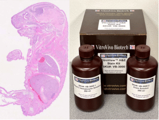
VitroView™ Hematoxylin and Eosin Stain Kit (250 ml per bottle)
SKU: VB-3000 $99.00 -
VitroView™ Hematoxylin and Eosin Stain Kit (500 ml per bottle)
SKU: VB-3000L $149.00 -
VitroView™ Hematoxylin and Eosin Stain Kit (30 ml per Dropper bottle)
SKU: VB-3000s $70.00 -
VitroView™ Mammary Gland Whole Mount Stain Kit
SKU: VB-3001 $219.00 -
VitroView™ Alcian Blue Hematoxylin-Orange G Stain Kit
SKU: VB-3002 $259.00 -
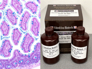
VitroView™ Alcian Blue Stain Kit
SKU: VB-3003 $195.00 -
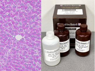
VitroView™ PAS Stain Kit
SKU: VB-3004 $245.00 -
VitroView™ Alcian Blue – PAS Stain Kit
SKU: VB-3005 $295.00 -
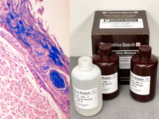
VitroView™ Luxol Fast Blue Stain Kit
SKU: VB-3006 $225.00 -
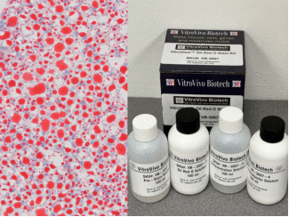
VitroView™ Oil Red O Stain Kit
SKU: VB-3007 $219.00 -
VitroView™ Alizarin Red Stain Kit
SKU: VB-3008 $95.00 -
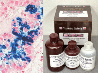
VitroView™ Prussian Blue Stain Kit
SKU: VB-3009 $195.00 -
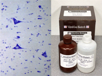
VitroView™ Nissl Stain Kit
SKU: VB-3010 $195.00 -
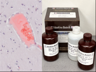
VitroView™ Congo Red Amyloid Stain Kit
SKU: VB-3011 $195.00 -
VitroView™ Sudan Black B Lipid Stain Kit
SKU: VB-3012 $215.00 -
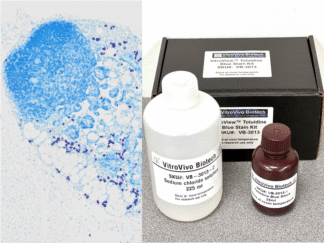
VitroView™ Toluidine Blue Stain Kit
SKU: VB-3013 $125.00 -
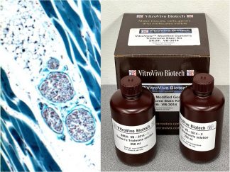
VitroView™ Modified Gomori’s Trichrome Stain Kit
SKU: VB-3014 $355.00 -
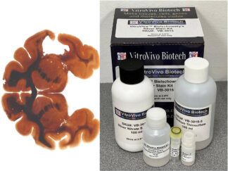
VitroView™ Bielschowsky’s Silver Stain Kit
SKU: VB-3015 $295.00 -
VitroView™ Masson’s Trichrome Stain Kit
SKU: VB-3016 $369.00 -
VitroView™ Picro-Sirius Red Stain Kit
SKU: VB-3017 $199.00 -
VitroView™ Reticulum Stain Kit
SKU: VB-3018 $335.00 -
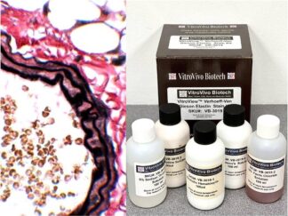
VitroView™ Verhoeff Van Gieson Elastin Stain Kit
SKU: VB-3019 $215.00 -
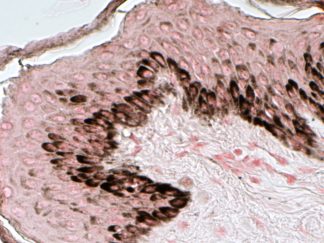
VitroView™ Fontana-Masson Stain Kit
SKU: VB-3020 $299.00 -
VitroView™ Rhodanine Copper Stain Kit
SKU: VB-3021 $185.00 -
VitroView™ Rhodanine Copper Stain Kit (small size)
SKU: VB-3021s $115.00 -
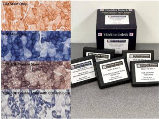
VitroView™ COX-SDH Double Histochemistry Stain Kit (For 50-100 Slides)
SKU: VB-3022 $495.00 -
VitroView™ COX Histochemistry Stain Kit (For 50-100 Slides)
SKU: VB-3022C $260.00 -
VitroView™ COX-SDH Double Histochemistry Stain Kit (For 10-20 Slides)
SKU: VB-3022s $195.00 -
VitroView™ Movat Pentachrome Stain Kit
SKU: VB-3023 $525.00 -
VitroView™ Southgate’s Mucicarmine Stain Kit
SKU: VB-3024 $299.00 -
VitroView™ Jones Stain Kit (Basement Membrane)
SKU: VB-3026 $320.00 -
VitroView™ Colloidal Iron Stain Kit
SKU: VB-3027 $288.00 -
VitroView™ Safranin O Stain Kit for Cartilage
SKU: VB-3028 $239.00 -
VitroView™ NADH-TR Histochemistry Stain Kit (For 50~100 slides)
SKU: VB-3029 $399.00 -
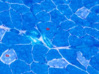
VitroView™ Metachromatic Myofibrillar ATPase Stain Kit
SKU: VB-3030 $459.00 -
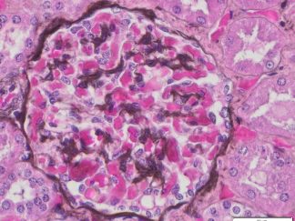
VitroView™ Periodic Acid Silver Methenamin Stain Kit
SKU: VB-3033 $375.00 -
VitroView™ Verhoeff Elastic Masson Trichrome Stain Kit
SKU: VB-3034 $425.00 -
VitroView™ Thioflavin S Stain Kit
SKU: VB-3035 $199.00 -
VitroView™ Martius Scarlet Blue (MSB) Stain Kit
SKU: VB-3036 $435.00 -
VitroView™ Gallyas Silver Stain Kit
SKU: VB-3037 $488.00 -
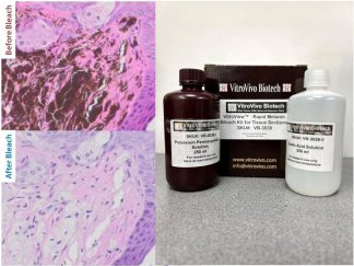
VitroView™ Rapid Melanin Bleach Kit for Tissue Sections
SKU: VB-3038 $109.00 -
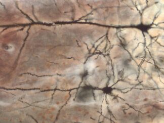
VitroView™ Golgi-Cox Stain Kit
SKU: VB-3039 $420.00 -
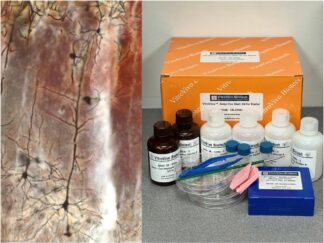
VitroView™ Golgi-Cox Stain Kit for Starter
SKU: VB-3039s $390.00 -
VitroView™ Nile Red Stain Kit
SKU: VB-3042 $228.00 -

VitroView™ Von Kossa Calcium Stain Kit
SKU: VB-3043 $218.00 -
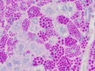
VitroView™ Combined Eosinophil-Mast Cell Stain Kit
SKU: VB-3044 $168.00 -
VitroView™ PAS-Diastase (PAS-D) Stain Kit
SKU: VB-3045 $386.00
Showing all 47 results