Description
Aluminum is believed to form a chelation complex with the carmine, changing the molecule to a positive charge allowing it to bind with the acid substrates of low density such as mucins. To stain mucin which is a secretion produced by a variety of epithelial cells and connective tissue cells. Excess mucin is secreted by epithelial cells in certain inflammations and in certain intestinal carcinomas. The mucicarmine technique is also useful in determining the site of a primary tumor in that finding mucin positive tumor cells in an area that does not contain mucin producing cells would indicate the tumor did not arise from that area. It is also useful in staining encapsulated fungi and cryptococcus.
Kit Components
| SKU# | Reagent Name | Volume (ml) |
| VB-3024-1 | Mayer’s Hematoxylin Solution | 250 |
| VB-3024-2 | Mucicarmine Solution | 250 |
| VB-3024-3 | Metanil yellow Solution | 250 |
Storage
Room temperature.
Protocol
- Deparaffinize in xylene I for 6 minutes and II for 6 minutes.
- Rehydrate: ethanol 100% (2 minutes)x2; ethanol 95% (2 minutes)x2; ethanol 70% (2 minutes)x1; Rinse in distilled water (5 minutes).
- Place in Mayer’s Hematoxylin Solution for 5 minutes.
- Wash in running tap water, 5 minutes.
- Incubate in mucicarmine solution at room temperature for 1 hour or microwave Hi power, 45 seconds.
- Rinse quickly in distilled water.
- Metanil yellow, 30 seconds to 1 minute.
- Dehydrate with 2 changes of 95% Ethanol and 2 changes of 100% Ethanol (2 minute per change).
- Clear with 3 changes of xylene (5 minute per change)
- Mount cover slip onto glass slide with Permount or some other suitable organic mounting medium.
Expected results
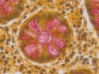
| Mucin | deep rose |
| Nuclei | black |
| Other tissue | elements yellow |
Control Tissue
Intestine or Colon.
Note
This product is intended for research purposes only. This product is not intended to be used for therapeutic or diagnostic purposes in humans or animals.
Precautions
Handle with care. Avoid contact with eyes, skin and clothing. Do not ingest. Wear gloves.
User Manual and Material Safety Data Sheet (MSDS) (PDF)
More Images


Frequently Asked Questions
What does this kit stain?
Mucin, a secretion produced by a variety of epithelial cells and connective tissue cells. Excess mucin is secreted by epithelial cells in certain inflammations and in certain intestinal carcinomas, and finding mucin-positive tumor cells in an area that does not contain mucin-producing cells can help determine the site of a primary tumor. The technique is also useful for staining encapsulated fungi and cryptococcus.
What do the results look like?
Mucin stains deep rose, nuclei stain black, and other tissue elements stain yellow.
What is in the kit?
Three reagents, 250 ml each: Mayer’s Hematoxylin Solution (VB-3024-1), Mucicarmine Solution (VB-3024-2), and Metanil Yellow Solution (VB-3024-3).
How should I store it?
At room temperature.
What are the key steps in the protocol?
After deparaffinizing in xylene and rehydrating through graded ethanols, sections are placed in Mayer’s Hematoxylin Solution for 5 minutes, then incubated in mucicarmine solution at room temperature for 1 hour or in a microwave on high power for 45 seconds, rinsed, and treated with Metanil yellow for 30 seconds to 1 minute, then dehydrated, cleared in xylene, and mounted with Permount or another suitable organic mounting medium.
What sample types and controls is it used with?
The protocol begins with deparaffinization in xylene, so it is designed for paraffin sections. The recommended control tissue is intestine or colon.