Description
The VitroView™ Senescence-Associated β-Galactosidase (SA-β-gal) Staining Kit is a sensitive and convenient histochemical staining system for the detection of senescence-associated β-galactosidase (SA-β-gal) activity, a widely used biomarker for identifying senescent cells.
Cellular senescence is associated with characteristic phenotypic changes, including increased cell size, flattened morphology, and alterations in gene expression. SA-β-gal activity detected at pH 6.0 is a commonly used histochemical indicator of the senescent phenotype and provides a convenient method for identifying senescent cells in research samples.
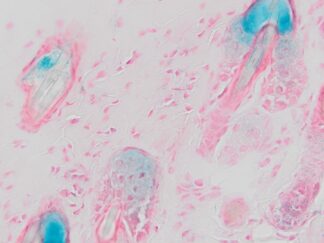
The kit is optimized for reliable and reproducible detection of SA-β-gal activity in cultured cells and frozen tissue sections. The resulting blue staining provides a straightforward visual method for identifying SA-β-gal-positive cells in studies of cellular aging, cancer biology, tissue aging, developmental biology, and drug discovery.
Applications
- Histochemical detection of SA-β-gal activity in frozen tissue sections
- Detection and visualization of senescent cells in cultured cells
- Research on cellular senescence and aging
- Cancer and tumor biology research
- Drug discovery and senescence-inducing treatment studies
- Evaluation of senescence-associated changes in experimental tissues and cells
Sample Compatibility
- Cell smears
- Cultured cells
- Frozen tissue sections
Kit Contents
| VB-3049-1 | Fixative Solution | 30 ml |
| VB-3049-2 | X-Gal (20x) | 1.5ml |
| VB-3049-3 | Staining Solution | 30ml |
| VB-3049-4 | Nuclear Fast Red Solution | 30 ml |
Storage
Store nuclear fast red solution at room temperature. Store other reagents at -20°C.
Procedure
1. Sample Preparation:
- For Adherent Cells / Coverslips: Discard the culture medium completely. Wash 3–4 times with PBS. Fix in fixative solution for 15–30 minutes. Wash 3 times with PBS. Proceed to Procedure Step 2.
- For Frozen Sections: Warm the frozen sections to room temperature. Fix in fixative solution for 15–30 minutes. Wash 3 times with PBS. Proceed to Procedure Step 2.
2. Staining
- Prepare staining working solution: add X-Gal (20x) to the staining solution to make 1x working staining solution and mix thoroughly.
- Use a hydrophobic barrier pen to draw a water-repellent circle around tissue sections or cells on the slide.
- Gently drop the working solution to cover the cells or tissue section on the glass slides and incubate at 37 °C for 1 -12 hours.
- (Optional) Nuclear Fast Red Counterstaining: For cultured cells, nuclear counterstaining with Nuclear Fast Red Solution is generally not necessary. For frozen tissue sections, Nuclear Fast Red counterstaining may be used to improve nuclear visualization and provide better tissue contrast. If desired, apply Nuclear Fast Red Solution directly to the sections and incubate for 2–5 minutes. Rinse the sections thoroughly with running tap water, followed by one rinse with deionized (DI) water. Proceed with mounting using an appropriate aqueous mounting medium.
3. Mounting: After completion of staining and washing, mount the stained slides using an aqueous, non-fluorescing mounting medium. For optimal results, VitroView™ Aqueous Mounting Medium with Coverslip Sealant Kit (SKU# VB-8004cs) is recommended. Seal edges with Coverslip Sealant if needed for long-term storage.
Expected Results under Bright-field Microscopy
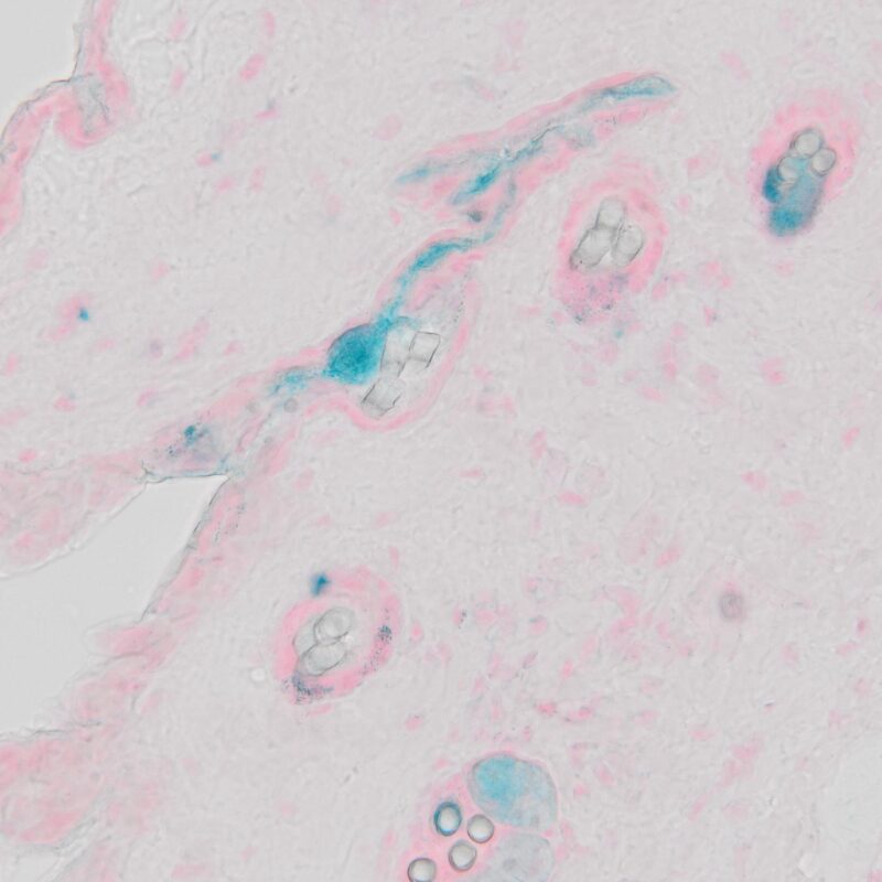
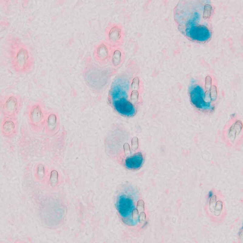
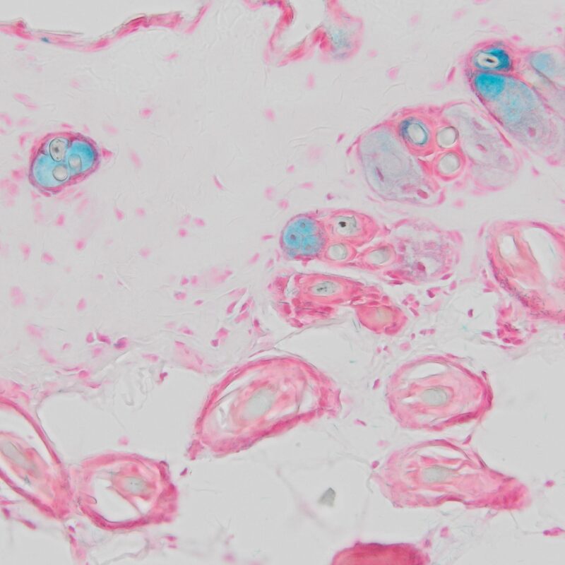
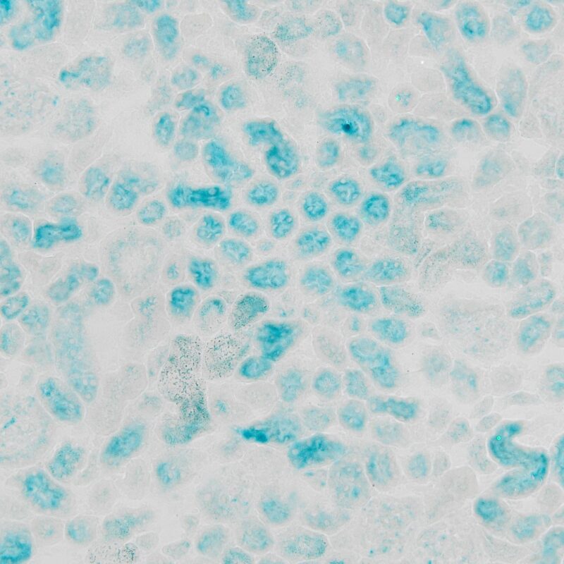
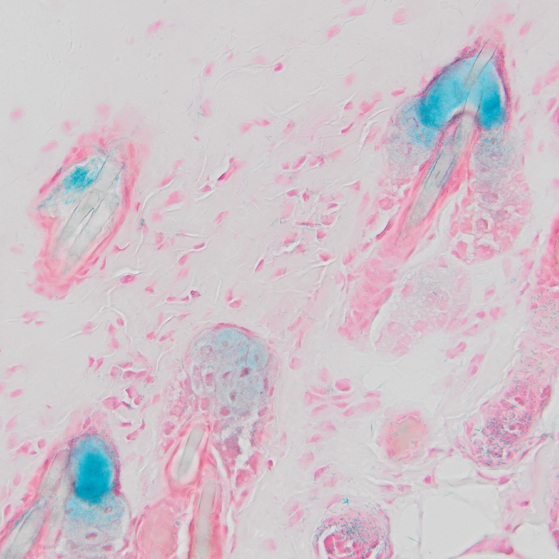
- Positive ———————blue
- Nuclei———————– red
Positive Controls
- Naturally Aged Mouse Tissue:Frozen sections of kidney or lung from aged mice (typically >12-24 months old) exhibit high levels of spontaneous cellular senescence and consistently produce a strong positive (blue) signal.
- Drug-Induced Senescence Tissue:Tissues collected from mice treated with DNA-damaging agents, such as bleomycin or doxorubicin, develop rapid, widespread senescence and provide reliable positive control.
- Senescent Cell Pellet (Cell-Based Positive Control):Primary human fibroblasts (e.g., WI-38 or IMR-90) induced into senescence via replicative exhaustion (high passage number) or oxidative stress (H2O2 treatment). Cell pellets can be prepared as frozen sections or cytospins.
Troubleshooting Guide
- Problem: High background – entire tissue section shows uniform faint blue staining (False positive)
Possible Causes & Solutions:
- Over-incubation:Incubation exceeding the recommended time (>24 hours) or temperature above 37°C can cause non-specific staining. Incubate strictly at 37°C without CO2 and do not exceed 16-24 hours.
- Inadequate washing:Residual fixative or endogenous factors can cause background. Ensure thorough washing with PBS after fixation and before adding the staining solution.
2. Problem: No signal detected in known positive control
Possible Causes & Solutions:
- Over-fixation:Prolonged fixation inactivates endogenous β-galactosidase. Reduce fixation time to 10-15 minutes for cells and 15-20 minutes for tissue sections.
- X-gal degradation:X-gal substrate is highly light-sensitive and unstable when improperly stored. Ensure the stock solution in DMSO is stored at -20°C, protected from light, and is fully dissolved without precipitate before use.
- Incorrect incubation temperature:SA-β-gal activity is temperature-sensitive. Confirm that the incubator maintains a steady 37°C, avoid temperature fluctuations, and incubate without CO2.
References
- Zhang W, Cheung TH. 2025. Detection of Cellular Senescence on Murine Muscle Tissue Sections by Senescence-Associated β-Galactosidase Staining. Methods in Molecular Biology, 2939:83–88
- Brandon M. Hall, et al, (2016). Aging of mice is associated with p16(Ink4a)- and β-galactosidase-positive macrophage accumulation that can be induced in young mice by senescent cells, 9 (8), 1867—1884
Note
This product is intended strictly for laboratory research purposes. It is not approved for therapeutic, clinical, or diagnostic procedures in humans or animals.
Precautions
Handle with care. Avoid contact with eyes, skin, and clothing. Do not ingest. Wear gloves.
More Images
|
|
|
|
|
|
|