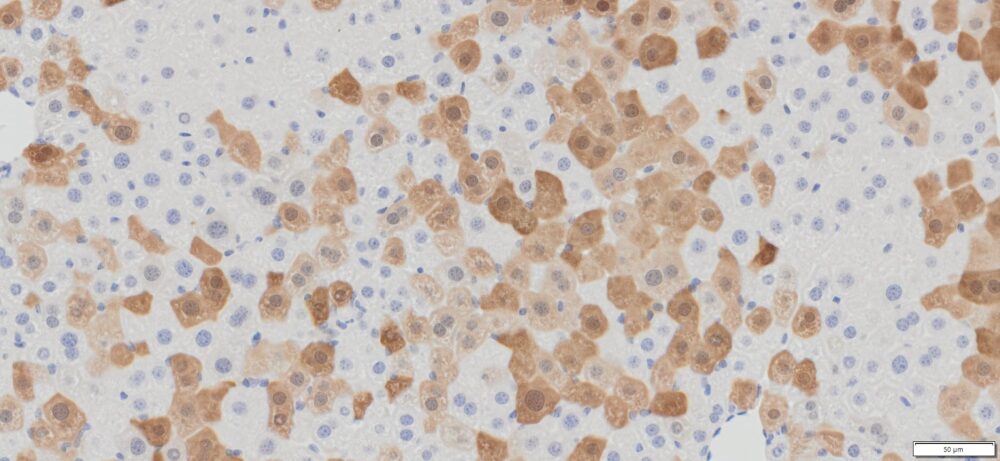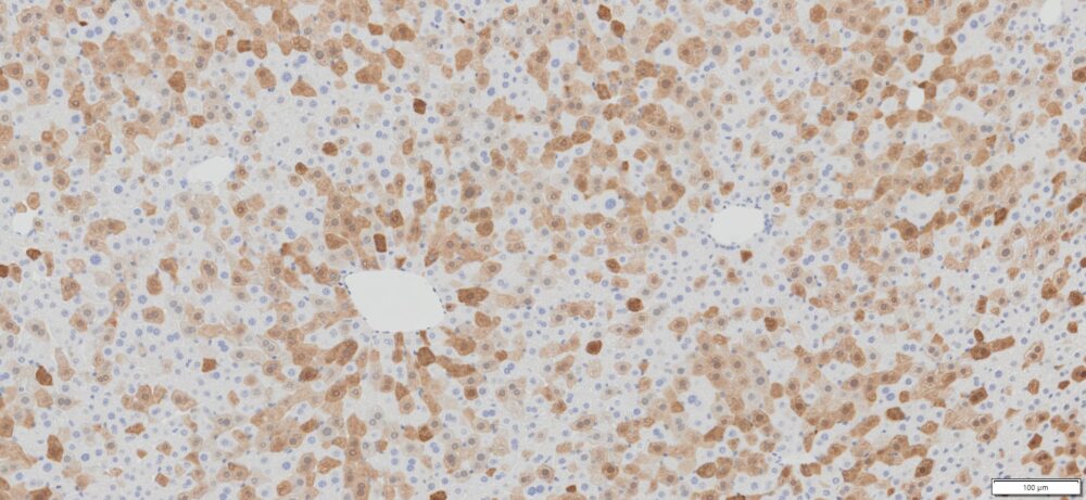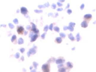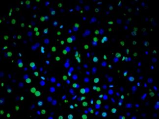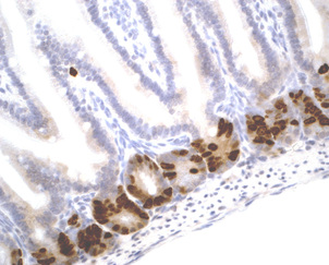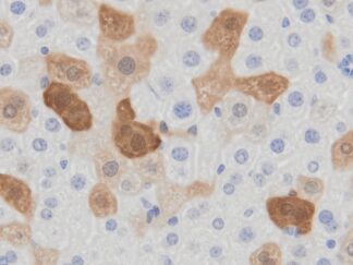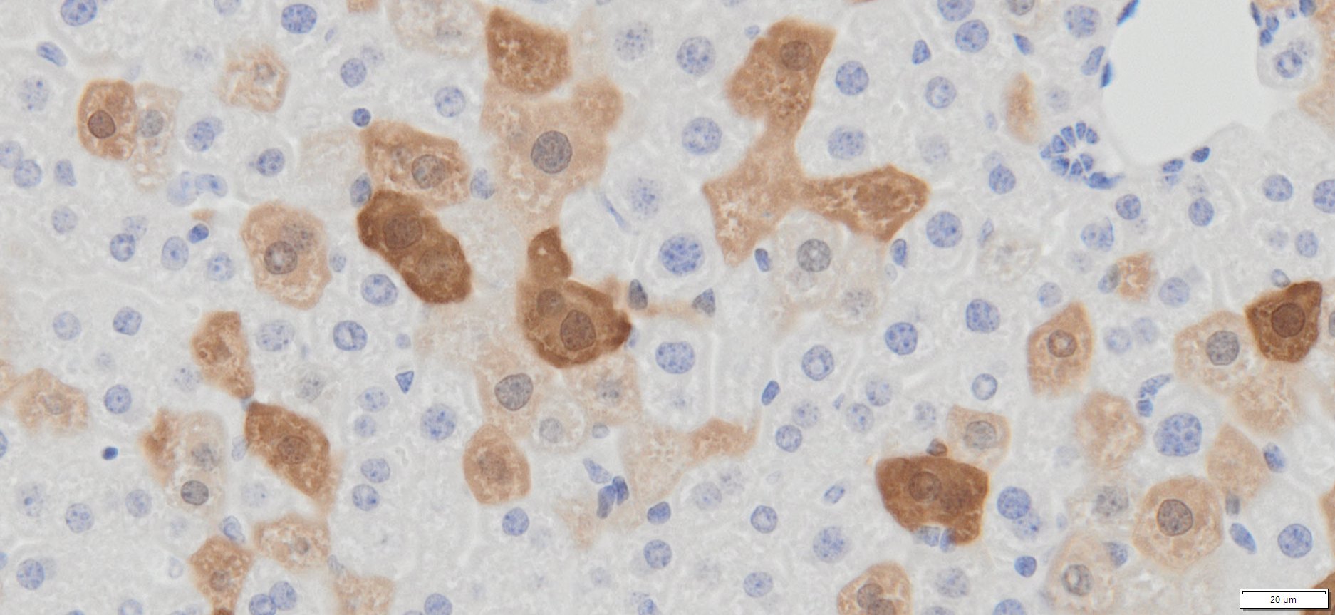
Description
Green fluorescent protein (GFP) is a 27 kDa monomeric protein that autocatalytically forms a fluorescent pigment. GFP has been used widely as a reporter protein for gene expression in eukaryotic and prokaryotic organisms, and GFP technology has revealed considerable new insights into the physiological activities of living cells. VitroView™ In Situ GFP Detection IHC Kit is a high-sensitivity immunohistochemistry (IHC) kit designed for the chromogenic detection of GFP and GFP fusion proteins in tissue sections and cultured cells. Using high-affinity anti-GFP antibodies and an optimized enzyme-based detection system, the kit enables precise localization of GFP-tagged proteins while preserving tissue morphology and histological architecture.
Native GFP fluorescence is often reduced or completely lost during formalin fixation, paraffin embedding, antigen retrieval, or long-term sample storage. The VitroView™ In Situ GFP Detection IHC Kit overcomes these limitations by converting GFP expression into a permanent chromogenic signal that can be visualized using a standard brightfield microscope. This approach provides superior sensitivity, enhanced signal stability, and long-term slide preservation compared with direct fluorescence detection.
The kit is optimized for formalin-fixed paraffin-embedded (FFPE) tissues, frozen sections, tissue microarrays, and cultured cells, producing strong, specific staining with minimal background. It is suitable for detecting GFP reporter genes, transgene expression, viral vector delivery, and GFP-tagged recombinant proteins in a wide variety of research applications.
Applications
- Detection of GFP-expressing cells in FFPE and frozen tissues
- Immunohistochemical analysis of GFP reporter gene expression
- Validation of transgenic animal models
- Evaluation of viral vector–mediated gene delivery
- Protein localization and tissue distribution studies
- Cancer, neuroscience, developmental biology, and stem cell research
Key Features
- High-sensitivity chromogenic detection of GFP and GFP fusion proteins
- Optimized for FFPE tissues, frozen sections, tissue microarrays, and cultured cells
- Permanent DAB staining for brightfield microscopy
- High specificity with low background staining
- Excellent tissue morphology preservation
- Robust, reproducible staining with optimized reagents and protocols
- Compatible with manual IHC staining platforms
Contents
| VB-4100-1 | 10×Ag Retrieval Solution | 50 ml |
| VB-4100-2 | RTU Blocking Buffer | 5 ml |
| VB-4100-3 | RTU anti-GFP Antibody | 5 ml |
| VB-4100-4 | RTU Polymeric Peroxidase Conjugated Secondary Antibody | 5 ml |
| VB-4100-5 | GFP Positive Control FFPE slides | 2 slides |
Note: RTU=ready-to-use
Reagents and Material Required but Not Provided
- Xylene and ethanol
- Distilled or deionized water
- 30% hydrogen peroxide
- 10 mM phosphate-buffered saline (PBS), pH 7.4
- Triton X-100
- Mini PAP Pen
- DAB Substrate Kit (Cat#: VB-6003 or VB-6003E)
- Hematoxylin (Cat#:VB-6004)
- Mounting Media
Storage Condition
Store at 2-8°C
Protocol
- Preparation of Slides
A. Cell Lines
- Grow cultured cells on sterile glass coverslips or slides overnight at 37 º C.
- Wash briefly with PBS.
- Fix as desired. Possible procedures include: a. 20 minutes with 10% formalin in PBS (keep wet); b. minutes with ice-cold methanol, allow air to dry; c.10 minutes with ice-cold acetone, allow air dry.
- Wash in PBS.
B. Frozen Sections
- Snap frozen fresh tissues in liquid nitrogen or isopentane pre-cooled in liquid nitrogen, embedded in OCT compound in cryomolds. Store the frozen tissue block at -80°C until ready for sectioning.
- Transfer the frozen tissue block to a cryotome cryostat (e.g., -20°C) prior to sectioning and allow the temperature of the frozen tissue block to equilibrate to the temperature of the cryotome cryostat.
- Section the frozen tissue block into a desired thickness (typically 5-10 µm) using the cryotome.
- Place the tissue sections onto glass slides suitable for immunohistochemistry (e.g. Superfrost).
- Sections can be stored in a sealed slide box at -80°C for later use.
- Before staining, warm slides at room temperature for 30 minutes and fix in ice-cold acetone or ice-cold methanol for 10 minutes. Air dry for 30 minutes.
- Wash in PBS
C. Paraffin Sections
- Deparaffinize sections in xylene, 3×5min.
- Hydrate with 100% ethanol, 2×2min.
- Hydrate with 95% ethanol, 2×2min.
- Rinse in distilled water.
- Follow procedure for pretreatment as required.
- Antigen retrieval
Prepare 100 ml of 1× Ag Retrieval working solution by mixing 10 ml of 10× Ag Retrieval Solution with 90 ml of ddH2O. Place the slides in a Coplin jar containing the 1× Ag Retrieval working solution and heat in a microwave oven for 3 min to bring the slides to a boil. Other sources of heat, such as a pressure cooker or a hot water bath, may be used. Maintain at a sub-boiling temperature for 10 minutes. Slowly cool the slides on the benchtop for 30 minutes.
Note: Do not use this pretreatment with frozen sections or cultured cells that are not paraffin-embedded.
- Staining Procedure
- Wash: Rinse sections in PBS-Triton X-100 (0.025%) for 2 × 2 minutes.
- Serum Blocking: Incubate sections with 3–4 drops of Ready-to-Use (RTU) normal goat serum for 30 minutes at room temperature.
- Primary Antibody: Incubate sections with RTU anti-GFP antibody for 1–2 hours at room temperature, or overnight at 4°C. Rinse sections in PBS for 2 × 2 minutes.
- Peroxidase Blocking (Optional): Incubate sections in 0.3% hydrogen peroxide ((text{H}_2text{O}_2)) in PBS for 10 minutes at room temperature, then rinse in PBS.
- Detection: Incubate sections with 3–4 drops of RTU polymeric peroxidase-conjugated secondary antibody for 30 minutes at room temperature.
- Wash: Rinse sections in PBS for 3 × 2 minutes.
- Chromogen / Substrate: Incubate sections with 3 drops of DAB working solution for 2–8 minutes. Monitor signal development under a microscope. Stop the reaction by rinsing in distilled water for 2 × 2 minutes. Note: Prepare DAB working solution by mixing 25 µL of DAB stock solution with 1 mL of DAB buffer (VB-6003).
- Counterstain: Incubate sections with 3 drops of RTU hematoxylin solution for 1–2 minutes.
- Wash: Rinse sections in running tap water for 2 × 2 minutes.
- Dehydration & Clearing: Dehydrate sections through graded ethanol series: 75% ethanol for 2 minutes, 95% ethanol for 2 minutes, and 100% ethanol for 2 × 3 minutes. Clear sections in xylene for 2 × 5 minutes.
- Mounting: Coverslip sections using an appropriate mounting medium
IHC Troubleshooting
High background staining
| Possible Cause | Solution |
| Endogenous peroxidase activity was incompletely blocked. | Incubate sections in 0.3% hydrogen peroxide in methanol or PBS for 10-30 minutes at room temperature. |
| Deparaffinization was incomplete. | Prepare new sections and deparaffinize according to standard laboratory protocol using fresh xylene or xylene substitute. |
| Inadequate rinsing of slides. | Gently rinse the slide with the wash buffer bottle and place it in the wash bath for 5 minutes. Gentle agitation of the wash bath may increase effectiveness. |
| Over-development of substrate. | Reduce incubation time. |
| Dehydration of specimen during staining. | Keep section wet. |
Negative staining on positive slides
| Possible Cause | Solution |
| Steps in the staining protocol were performed in incorrect sequence. | Repeat the procedure. |
| Primary or secondary antibody incubation steps were omitted. | Repeat the procedure. |
| Labile antigens were destroyed. | Use fresh cutting slides. Use paraffin wax with a melting temperature of 55- 58ºC. Wax used for embedding should not exceed 60 ºC. |
| Specimen was improperly fixed and/or processed. | Check manufacturer’s specifications regarding recommended fixative. |
| Specimen dehydrates during staining. | Repeat the procedure by following the manufacturer’s protocol. |
Weak staining on all slides
| Possible Cause | Solution |
| Specimen retained excess liquid after rinsing steps. | Remove excess liquid after rinsing steps. |
| Incubation times were insufficient. | Prolong incubation time. |
| Substrate prepared improperly. | Check compatibility of buffer ingredients with enzyme and substrate-chromogen reagents. Repeat staining. |
| Deparaffinization was incomplete (staining may be accompanied by high background). | Prepare new sections and de-paraffinize according to standard laboratory protocol using fresh xylene or xylene substitute. |
Warning
DAB is a possible carcinogen. Please take necessary precautions.
Note
This product is intended for research purposes only. This product is not intended to be used for therapeutic or diagnostic purposes in humans or animals. Avoid contact with eyes, skin and clothing. Do not ingest. Wear gloves.
User Manual (PDF)