Description
| Organ& Tissue | Pathology Diagnosis | Gender/Age | Tissue of Origin | % Tumor Area | Grade | TMN Stage | Biomarkers |
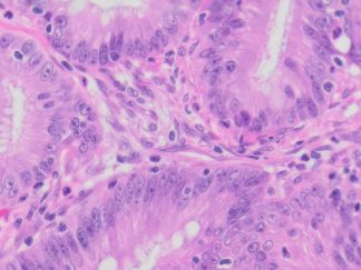
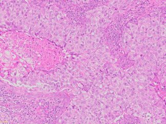
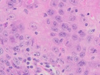
| Tongue | Well differentiated squamous cell carcinoma | Female/82 | Right hemiglossectomy | 60% | I | T1N0M0 |
Slide Preparation
Human tissue was fixed in formalin immediately after excision and embedded in paraffin. The tissue sections were 5 µm in thickness and mounted each on positively charged glass slides.
Sections/slide
1 section on each slide
Slides/Package
5 slides per package
Applications
Histology stain, IHC/IF, FISH and ISH
Storage Condition
Store at 2-8ºC
More Images of H&E Stain and IHC


Product Data Sheet and Material Safety Data Sheet (MSDS) (PDF)
Frequently Asked Questions
What diagnosis do these tongue sections show?
Well differentiated squamous cell carcinoma of the tongue, from a right hemiglossectomy specimen.
What donor is listed for this case?
An 82-year-old female donor.
What tumor area, grade, and stage are recorded?
Tumor area 60%, grade I, and TNM stage T1N0M0.
How thick are the sections, and how are they mounted?
The sections are 5 µm thick, cut from tissue fixed in formalin immediately after excision and embedded in paraffin, and mounted on positively charged glass slides, one section per slide.
How many slides come in a package?
5 slides per package.
What applications and storage conditions are listed?
The listed applications are histology staining, IHC/IF, FISH, and ISH, and the slides should be stored at 2–8 °C. H&E example images at 4x, 10x, and 20x are shown on the product page.