Description
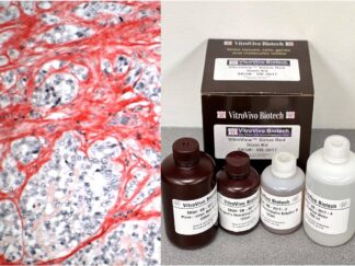
Mitochondria, often referred to as the “powerhouses” of the cell, play a crucial role in cellular energy production, metabolism, and various other cellular processes. Understanding mitochondrial function is critical to research areas such as cellular bioenergetics, neurodegenerative diseases, cancer, and aging. One key aspect of mitochondrial function is the activity of cytochrome c oxidase (COX), the terminal enzyme of the electron transport chain (ETC) involved in oxidative phosphorylation. COX is essential for the reduction of oxygen to water, which is coupled with the synthesis of ATP.
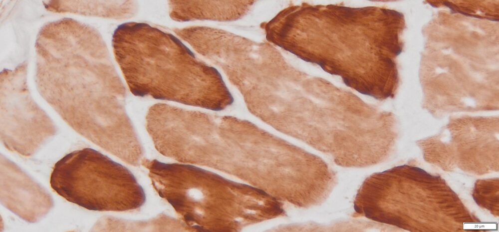
The COX Histochemistry Stain Kit is a valuable tool for researchers investigating mitochondrial function at the tissue and cellular level. This histochemical method allows for the direct visualization of COX activity within intact cells and tissues, providing a practical approach for studying mitochondrial bioenergetics.
Kit Components
| SKU# | Reagent | Size |
| VB-3022-1 | COX A Solution | 1ml×5 |
| VB-3022-2 | COX B Solution | 1ml×5 |
Storage
Store at -20°C
Protocol
- Tissue preparation for cryosectioning
- Euthanize the animal using cervical dislocation or decapitation, following the approved ethical permit.
- Rapidly collect tissues of interest without fixation and immediately freeze them on dry ice. For optimal morphology, tissues may require freezing in isopentane or propane chilled with liquid nitrogen.
- Wrap tissues in aluminum foil and store at -80 °C until sectioning.
- Embed frozen tissues in preparation for cryosectioning.
- Cut 10-14 μm cryostat sections, thaw them onto slides at room temperature for 2-5 minutes, and store slides without cover-slipping at -70 °C until use.
- Prepare COX Incubation Solution: Thaw one vial of COX A Solution (VB-3022-1) and one vial of COX B Solution (VB-3022-2). Mix one vial of COX A Solution with one vial of COX B Solution thoroughly.
- Rinse: Wash the slides with PBS to remove any residual OCT from the glass.
- Incubate with COX Solution: Immediately apply 80–200 µL of the COX incubation solution onto frozen sectioned slides in a humidity chamber. Incubate in the dark at room temperature for 1–1.5 hours.
- Check Staining: Assess the staining and extend the incubation time if needed.
- Rinse: Wash the slides in PBS.
- Dehydrate: Perform two changes of 95% ethanol followed by two changes of 100% ethanol, with each step lasting 2 minutes.
- Clear: Immerse the slides in three changes of xylene, with each step lasting 5 minutes.
- Mount Coverslip: Apply Permount or another suitable organic mounting medium and place a coverslip onto the glass slide.
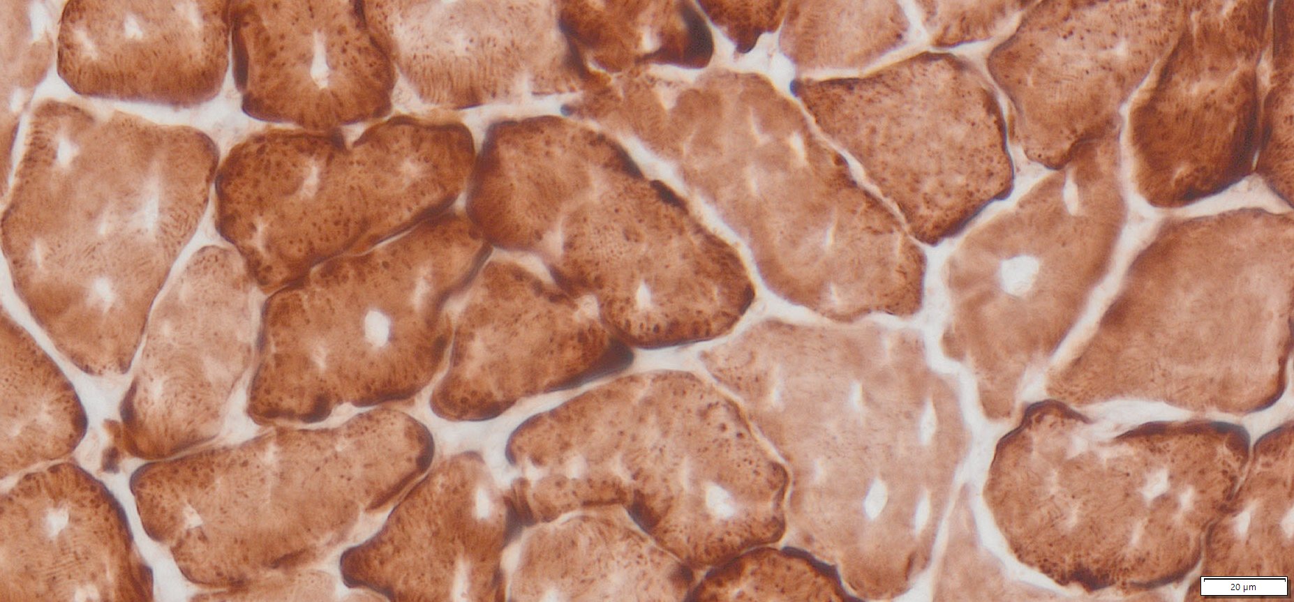
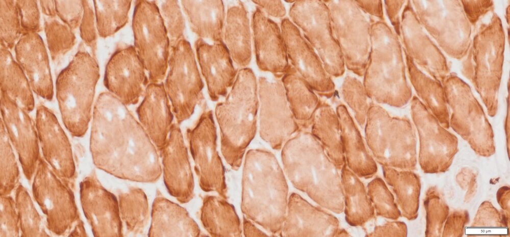
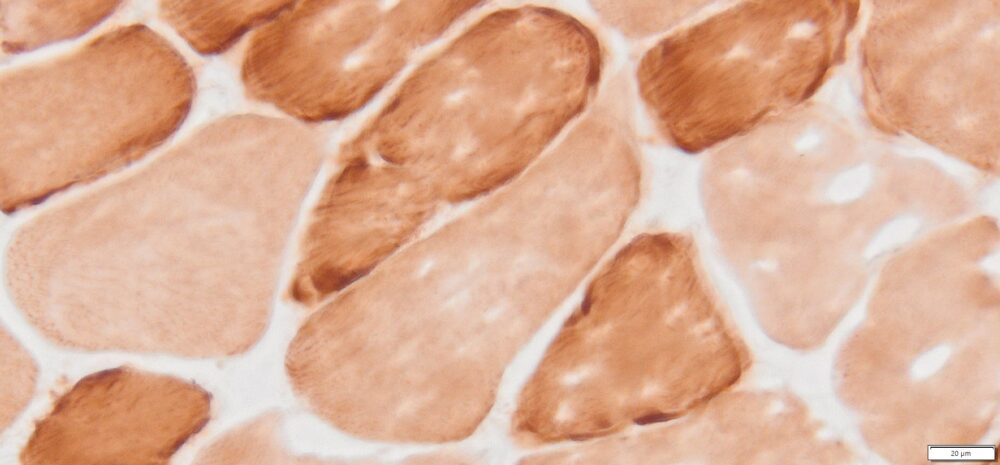
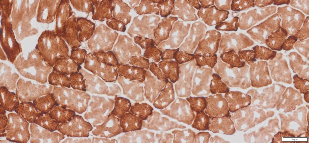
Results
| Cytochrome Oxidase positive mitochondria——- | Brown |
More Images
References
- Ross, J.M. Visualization of Mitochondrial Respiratory Function using Cytochrome C Oxidase / Succinate Dehydrogenase (COX/SDH) Double-labeling Histochemistry. J. Vis. Exp. (57), e3266, DOI : 10.3791/3266 (2011).
- Seligman etal (1968) J Cell Biol 38:1-14.
- Loughlin M. (1993). Muscle biopsy. A laboratory investigation. Butterworth-Heinemann p.38-39.
- Sheehan D, Hrapchak B. (1987). Histotechnology, 2nd Ed. Batelle Press, Columbus p306-307
Note
This product is intended for research purposes only. This product is not intended to be used for therapeutic or diagnostic purposes in humans or animals.
Precautions
Handle with care. Avoid contact with eyes, skin and clothing. Do not ingest. Wear gloves.
User Manual and Material Safety Data Sheet (MSDS) (PDF)
VB-3022 MSDS