Description
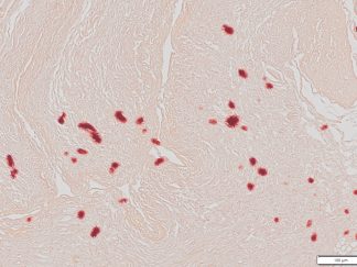
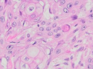
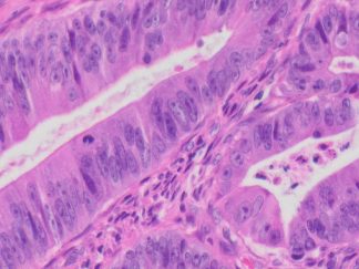
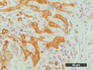
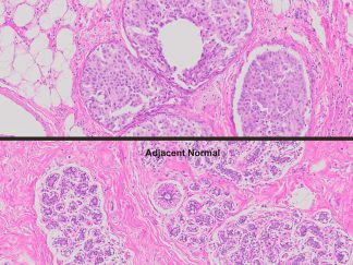
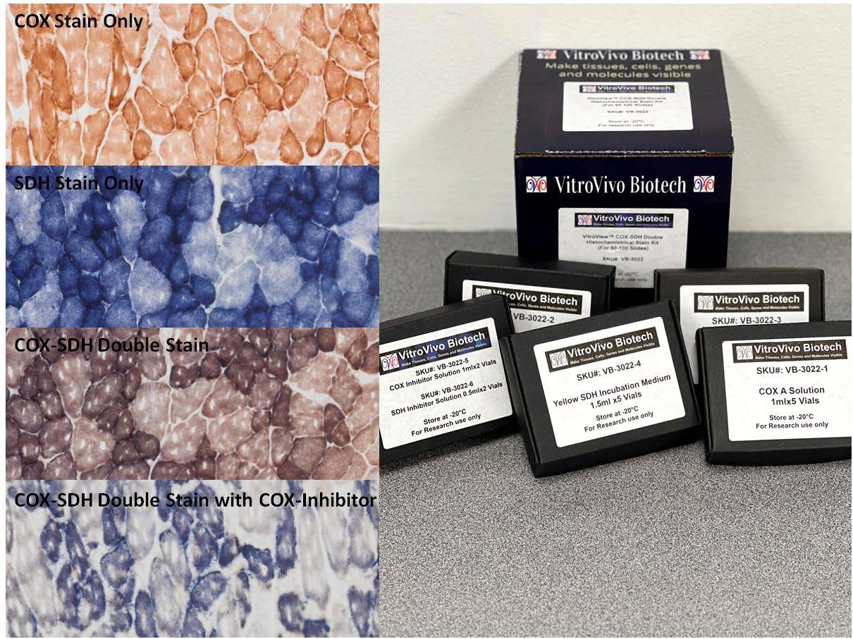
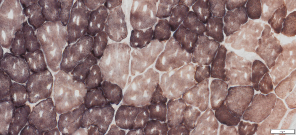
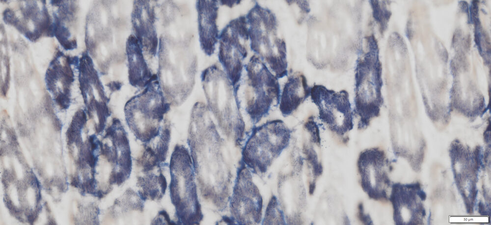
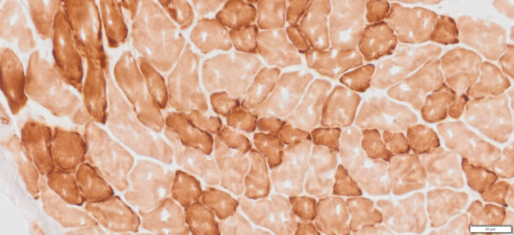
Observing respiratory enzyme activity provides a straightforward method for investigating mitochondrial dysfunction. Cytochrome c oxidase (COX) is crucial for mitochondrial function, while succinate dehydrogenase (SDH) is entirely encoded by nuclear DNA and remains unaffected by mitochondrial DNA (mtDNA) mutations. A combined COX/SDH stain highlights COX-negative fibers, which may appear SDH-positive and correspond to ragged red fibers. Since three of the 13 COX subunits are mtDNA-encoded, mtDNA mutations can impair COX activity, whereas SDH remains unaffected. In mitochondrial myopathies, ragged red fibers are typically COX-negative, except in Mitochondrial Encephalopathy, Lactic Acidosis, and Stroke-like Episodes (MELAS). Additionally, intra-fiber mosaicism—characterized by a mix of COX-deficient (bluish) and COX-positive (brownish) mitochondria within the same fiber—is well demonstrated with this staining technique. Mitochondrial diseases, aging, and age-related conditions often lead to cells with reduced or absent COX activity, making this method valuable for research and diagnostics.
Kit Introduction Viedeo
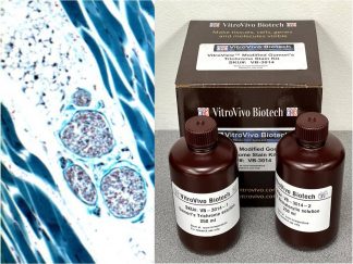
Kit Components
| SKU# | Reagent | Size |
| VB-3022-1 | COX A Solution | 1ml×5 |
| VB-3022-2 | COX B Solution | 1ml×5 |
| VB-3022-3 | Succinate Solution | 0.3ml×5 |
| VB-3022-4 | Yellow SDH Incubation Medium | 1.5 ml×5 |
| VB-3022-5 | COX Inhibitor Solution | 1ml×2 |
| VB-3022-6 | SDH Inhibitor Solution | 0.5 ml×2 |
Storage
Store at -20°C
Protocol
- Tissue preparation for cryosectioning
- Euthanize the animal using cervical dislocation or decapitation, following the approved ethical permit.
- Rapidly collect tissues of interest without fixation and immediately freeze them on dry ice. For optimal morphology, tissues may require freezing in isopentane or propane chilled with liquid nitrogen.
- Wrap tissues in aluminum foil and store at -80 °C until sectioning.
- Embed frozen tissues in preparation for cryosectioning.
- Cut 10-14 μm cryostat sections, thaw them onto slides at room temperature for 2-5 minutes, and store slides without cover-slipping at -70 °C until use.
- Prepare COX Incubation Solution: Thaw one vial of COX A Solution (VB-3022-1) and one vial of COX B Solution (VB-3022-2). Mix one vial of COX A Solution with one vial of COX B Solution thoroughly.
- Rinse: Wash the slides with PBS to remove any residual OCT from the glass.
- Incubate with COX Solution: Immediately apply 80–200 µL of the COX incubation solution onto frozen sectioned slides in a humidity chamber. Incubate in the dark at room temperature for 1–1.5 hours.
- Check Staining: Assess the staining and extend the incubation time if needed.
- Rinse: Wash the slides in PBS.
- Prepare Fresh SDH Incubation Medium: Thaw a 0.3 mL vial of Succinate Solution (VB-3022-3) and a 1.5 mL vial of Yellow SDH Incubation Medium (VB-3022-4). Mix 0.3 mL of Succinate Solution with 1.5 mL of Yellow SDH Incubation Medium, or combine them at a 1:5 ratio. Mix well before use.
- Incubate with SDH Solution: Apply 80–200 µL of the SDH incubation medium onto the slide and incubate at 37°C for 40 minutes.
- Dehydrate: Perform two changes of 95% ethanol followed by two changes of 100% ethanol, with each step lasting 2 minutes.
- Clear: Immerse the slides in three changes of xylene, with each step lasting 5 minutes.
- Mount Coverslip: Apply Permount or another suitable organic mounting medium and place a coverslip onto the glass slide.
Appropriate specificity controls
- Specificity Control for COX Activity: Before step 3, apply 80-200 µL of COX Inhibitor Solution (VB-2022-5) to the slide. Incubate for 5 minutes, then proceed to step 3.
- Specificity Control for SDH Activity: Prepare a solution by mixing SDH Inhibitor Solution (VB-3022-6) with Yellow SDH Incubation Medium (VB-3022-4) at a 1:5 ratio. Apply 80-200 µL of this mixture onto the slide, ensuring the section is covered, and incubate at 37°C for 40 minutes. This step replaces step 7.
Results
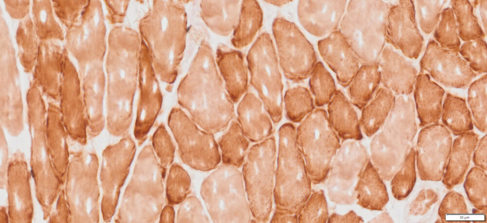
| Cytochrome Oxidase positive mitochondria——- | Brown |
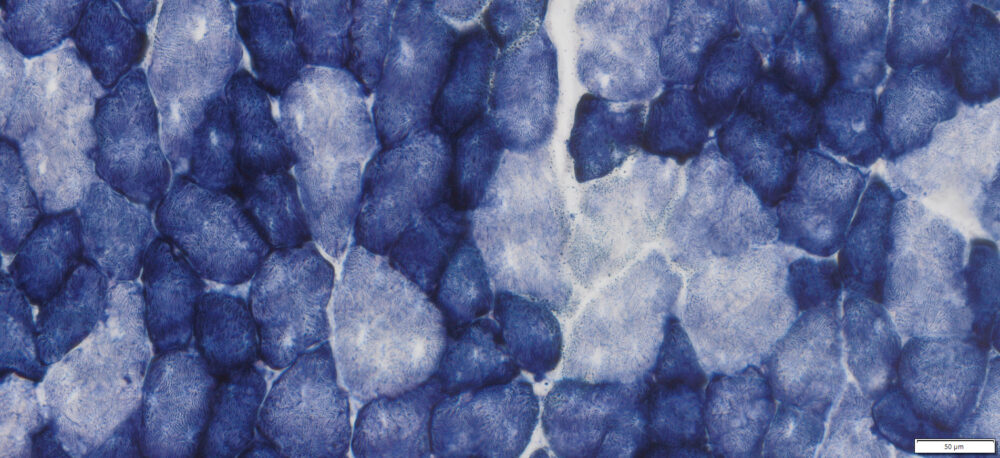
| Cytochrome Oxidase negative mitochondria——- | Blue |
More Images
References
- Ross, J.M. Visualization of Mitochondrial Respiratory Function using Cytochrome C Oxidase / Succinate Dehydrogenase (COX/SDH) Double-labeling Histochemistry. J. Vis. Exp. (57), e3266, DOI : 10.3791/3266 (2011).
- Seligman etal (1968) J Cell Biol 38:1-14.
- Loughlin M. (1993). Muscle biopsy. A laboratory investigation. Butterworth-Heinemann p.38-39.
- Sheehan D, Hrapchak B. (1987). Histotechnology, 2nd Ed. Batelle Press, Columbus p306-307
Recent Publications Using VB-3022
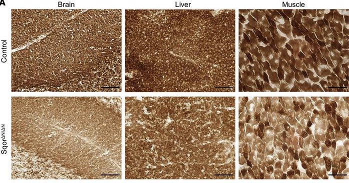
- Kanemaru Eiki et al. (2024). Exclusion of sulfide:quinone oxidoreductase from mitochondria causes Leigh-like disease in mice by impairing sulfide metabolism. J Clin Invest. 134(15):e170994.
2. Dombrecht D, Daele UV, Asbroeck BV, David R. Schieffelers DR, Guns PJ,Breda EV (2023). Skeletal muscle wasting after burn is regulated by a decrease in anabolic signaling in the early flow phase. BurnsVolume; 49 ( 7): 1574-1584
3. Dang X, Zhang L, Franco S, Dorn II GW (2023). Activating mitofusins interrupts mitochondrial degeneration and delays disease progression in SOD1 mutant amyotrophic lateral sclerosis. Human Molecular Genetics; 32 (7): 1208–1222
4. Mosteiro L, Nguyen TTT, Hankeova S, Alvarez-Sierra D, Reichelt M, Vandriel SM, Lai Z, Choudhury FK, Sangaraju D, Kamath DM, Scherl A, Pujol-Borrell R, Robert Piskol R & Christian W. Siebel (2023). Notch signaling in thyrocytes is essential for adult thyroid function and mammalian homeostasis. Nature Metabolism; 5: 2094–2110
Note
This product is intended for research purposes only. This product is not intended to be used for therapeutic or diagnostic purposes in humans or animals.
Precautions
Handle with care. Avoid contact with eyes, skin and clothing. Do not ingest. Wear gloves.