Description
Sample Basic Information
| Tissue ID | Organ | Pathology Diagnosis | Gender | Age | Grade | TMN | IHC Data | Sample Format |
| Hu-07002A | Human colon | Normal colon tissue | Male | 57 | N/A | N/A | Ki67 IHC | FFPE |
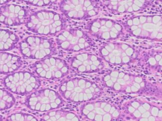
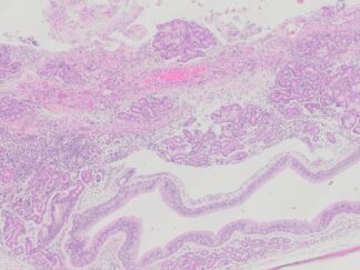
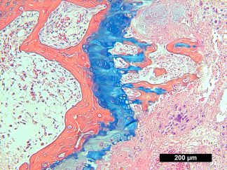
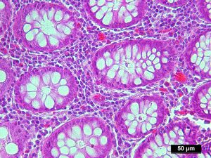
Tissue H&E Staining and IHC images
 H&E Stain H&E Stain |
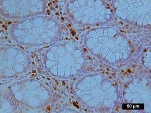 Ki67 IHC Ki67 IHC |
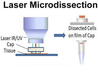
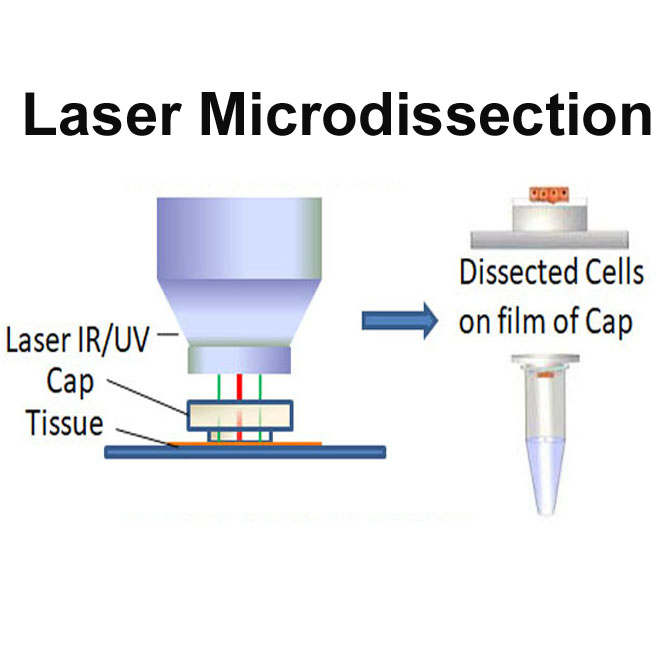
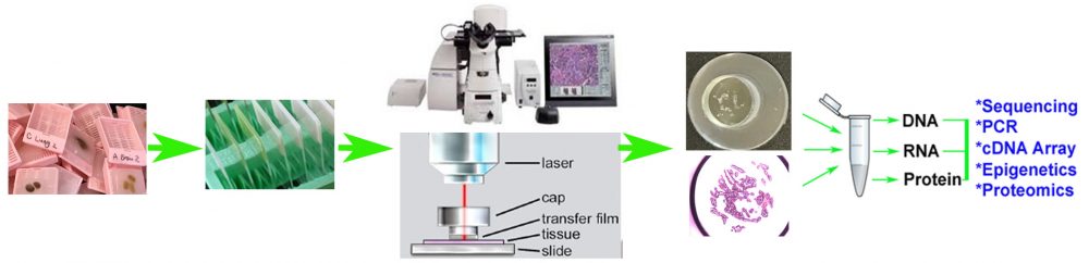
Flowchart of LCM for The Product Preparation
- FFPE samples are cut at 6 microns and mounted onto pen membrane glass slides and let the slides dry for 2 hours at room temperature.
- After deparaffinization and rehydration, quick H&E staining is performed.
- Arcturus XT Laser Microdissection System is used for LCM. Microdissection is performed using IR and UV laser and CapSure Macro LCM Cap.
- The total dissected cells may be varied between 10,000 to 50,000 cells which depend on tissue type.
- Protein is extracted from LCM Caps by following the protocol from Methods; 2016, 104: 154-162
Quality Control
- The tissue H&E staining slides are examined by certified pathologists. Pathological re-confirmation report is generated and digital image captured.
- LCM is supervised by our certified pathologist.
- Three example LCM images including Before, After LCM, and LCM cap images from LCM tissue slides are taken to demonstrate the dissected cells.
Extraction Protocol for Protein from LCM Caps
For protein extraction, you can follow the protocol from Longuespée R, et al. A laser microdissection-based workflow for FFPE tissue microproteomics: Important considerations for small sample processing, Methods;2016, 104: 154-162
Product Format and Size:
Protein in 50 μl of lysis buffer from 2 LCM caps.
Applications
The protein lysate can be used for proteomics.
Storage
Store at -80ºC
References
- Longuespée R, et al. A laser microdissection-based workflow for FFPE tissue microproteomics: Important considerations for small sample processing, Methods;2016, 104: 154-162
- Cai J, et al. The Use of Laser Microdissection in the Identification of Suitable Reference Genes for Normalization of Quantitative Real-Time PCR in Human FFPE Epithelial Ovarian Tissue Samples. PLoS ONE 2014, 9(4): e95974.
- Drummond ES, et al. Proteomic analysis of neurons microdissected from formalin-fixed, paraffin-embedded Alzheimer’s disease brain tissue.Sci Rep. 2015;5:15456
- Bockmeyer CL, et al. Recommendations for mRNA analysis of micro-dissected glomerular tufts from paraffin-embedded human kidney biopsy samples. BMC Mol Biol. 2018;19(1):2.
- Murray, Graeme I. (Ed.). Laser Capture Microdissection (Methods and Protocols), © 2018